. 2017 Jul 28;4(9):680-686.
doi: 10.1002/acn3.396. eCollection 2017 Sep.

Antibodies to TRIM46 are associated with paraneoplastic neurological syndromes

Affiliations

Antibodies to TRIM46 are associated with paraneoplastic neurological syndromes

Marleen H van Coevorden-Hameete et al. Ann Clin Transl Neurol. .

Abstract

Paraneoplastic neurological syndromes (PNS) are often characterized by the presence of antineuronal antibodies in patient serum or cerebrospinal fluid. The detection of antineuronal antibodies has proven to be a useful tool in PNS diagnosis and the search for an underlying tumor. Here, we describe three patients with autoantibodies to several epitopes of the axon initial segment protein tripartite motif 46 (TRIM46). We show that anti-TRIM46 antibodies are easy to detect in routine immunohistochemistry screening and can be confirmed by western blotting and cell-based assay. Anti-TRIM46 antibodies can occur in patients with diverse neurological syndromes and are associated with small-cell lung carcinoma.


Figures
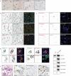
Figure 1
Figure 1
Identification and validation of TRIM46 as neuronal autoantigen. (A) Immunohistochemistry (IHC) of adult rat brains stained with the patients’ serum. The figures depict a part of the cortex showing prominent staining of the axon initial segment (AIS) (indicated with arrows) by the patients’ sera, which is absent in the staining with healthy control serum. Scale bars: 50 μm (B) IHC of P5 mouse cortex. The patients’ sera (green) stain the initial part of the axon and partially colocalize with the AIS marker ankyrinG (red). Scale bars: 20 μm. (C) Immunocytochemistry of cultured rat hippocampal neurons (DIV25). The patients’ sera (red) stain the initial part of the axon and partially colocalize with the AIS marker ankyrinG (green) but not with the dendritic marker MAP2 (blue). Scale bars: 20 μm. (D) HeLa cells expressing TRIM46‐GFP (green) were fixed, permeabilized and stained with patients’ or healthy control sera (red). The patients’ sera strongly recognize TRIM46, whereas the control serum does not. Scale bars: 10 μm. (E) Western blots using lysates of HEK cells overexpressing GFP or TRIM46‐GFP. The blots were stained with a GFP antibody, TRIM46 antibody, patients’ or healthy control sera. The patients’ sera recognize TRIM46‐GFP on blot but not GFP. (F) IHC of tumor tissue from patient 1, stained with hematoxylin‐eosin (HE), the lung carcinoma marker thyroid transcription factor 1 (TTF‐1), normal rabbit serum, and TRIM46 antibody. The picture shows TRIM46 expression in a subset of tumor cells. Stainings were performed on sequential slides, pictures were taken in the same area of the sample. Scale bars: 25 μm.
Figure 2
Figure 2
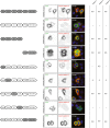
Epitope mapping of anti‐TRIM46 autoantibodies. Schematic representation of TRIM46 (gray) and TRIM36 (white). CC = coiled‐coil. COS = C‐terminal subgroup one signature. FN 3 = Fibronectin type III. GFP‐tagged truncated TRIM46 and chimeric proteins of TRIM46 and TRIM36 (green) expressed in HeLa cells and stained with patient's serum (red). The patients’ sera recognize the TRIM46 C‐terminus and N‐terminal B‐box, and CC region.

References

    1. Graus F, Delattre JY, Antoine JC, et al. Recommended diagnostic criteria for paraneoplastic neurological syndromes. J Neurol Neurosurg Psychiatry 2004;75:1135–1140. - PMC - PubMed
    1. Probst C, Komorowski L, de Graaff E, et al. Standardized test for anti‐Tr/DNER in patients with paraneoplastic cerebellar degeneration. Neurol Neuroimmunol Neuroinflamm 2015;2:e68. - PMC - PubMed
    1. de Graaff E, Maat P, Hulsenboom E, et al. Identification of delta/notch‐like epidermal growth factor‐related receptor as the Tr antigen in paraneoplastic cerebellar degeneration. Ann Neurol 2012;71:815–824. - PubMed
    1. Graus F, Keime‐Guibert F, Rene R, et al. Anti‐Hu‐associated paraneoplastic encephalomyelitis: analysis of 200 patients. Brain 2001;124(Pt 6):1138–1148. - PubMed
    1. Berghs S, Ferracci F, Maksimova E, et al. Autoimmunity to beta IV spectrin in paraneoplastic lower motor neuron syndrome. Proc Natl Acad Sci USA 2001;98:6945–6950. - PMC - PubMed