. 2017 Dec;21(12):3178-3189.
doi: 10.1111/jcmm.12871. Epub 2017 Sep 14.

Salidroside suppressing LPS-induced myocardial injury by inhibiting ROS-mediated PI3K/Akt/mTOR pathway in vitro and in vivo

Affiliations

Salidroside suppressing LPS-induced myocardial injury by inhibiting ROS-mediated PI3K/Akt/mTOR pathway in vitro and in vivo

Lvyi Chen et al. J Cell Mol Med. 2017 Dec.

Abstract

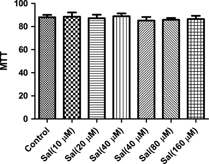
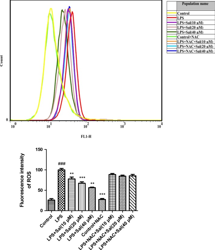
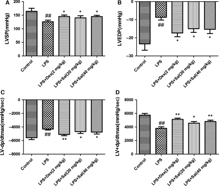
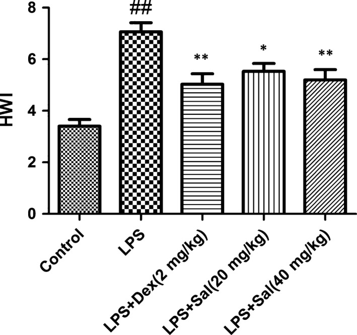
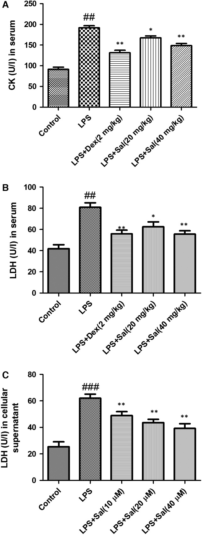
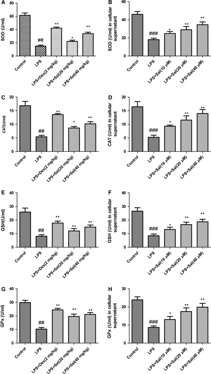
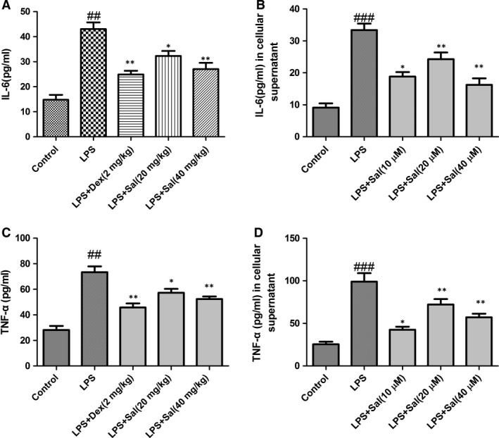
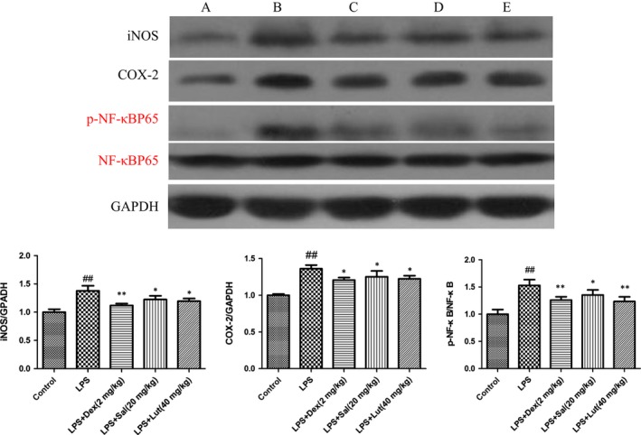
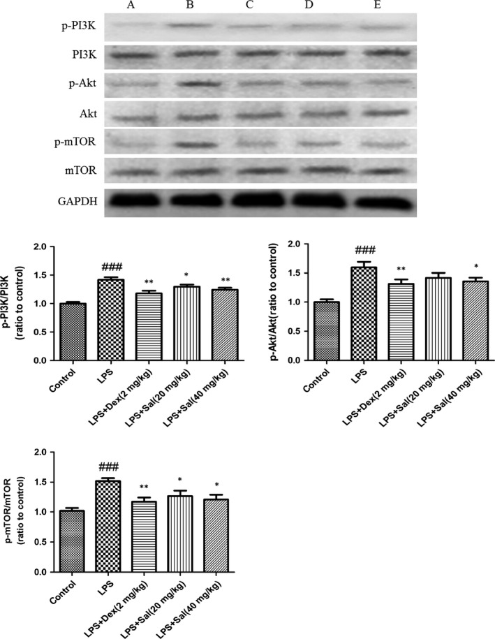
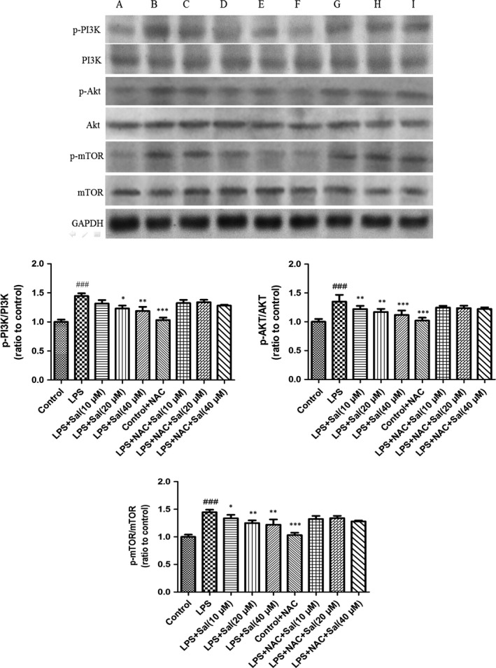
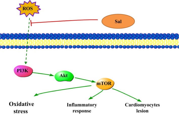
The purpose of the present study was to investigate the effect of salidroside (Sal) on myocardial injury in lipopolysaccharide (LPS)-induced endotoxemic in vitro and in vivo. SD rats were randomly divided into five groups: control group, LPS group (15 mg/kg), LPS plus dexamethasone (2 mg/kg), LPS plus Sal groups with different Sal doses (20, 40 mg/kg). Hemodynamic measurement and haematoxylin and eosin staining were performed. Serum levels of creatine kinase (CK), lactate dehydrogenase, the activities of the antioxidant enzymes catalase (CAT), superoxide dismutase (SOD), glutathione peroxidase (GSH-px), glutathione, tumour necrosis factor-α (TNF-α), interleukin-6 (IL-6), and interleukin-1β (IL-1β) were measured after the rats were killed. iNOS, COX-2, NF-κB and PI3K/Akt/mTOR pathway proteins were detected by Western blot. In vitro, we evaluated the protective effect of Sal on rat embryonic heart-derived myogenic cell line H9c2 induced by LPS. Reactive oxygen species (ROS) in H9c2 cells was measured by flow cytometry, and the activities of the antioxidant enzymes CAT, SOD, GSH-px, glutathione-S-transferase, TNF-α, IL-6 and IL-1β in cellular supernatant were measured. PI3K/Akt/mTOR signalling was examined by Western blot. As a result, Sal significantly attenuated the above indices. In addition, Sal exerts pronounced cardioprotective effect in rats subjected to LPS possibly through inhibiting the iNOS, COX-2, NF-κB and PI3K/Akt/mTOR pathway in vivo. Furthermore, the pharmacological effect of Sal associated with the ROS-mediated PI3K/Akt/mTOR pathway was proved by the use of ROS scavenger, N-acetyl-l-cysteine, in LPS-stimulated H9C2 cells. Our results indicated that Sal could be a potential therapeutic agent for the treatment of cardiovascular disease.

Keywords: LPS; ROS; H9C2; PI3K/Akt/mTOR; myocardial injury; salidroside.


Figures

Figure 1
Figure 1
Animal treatment protocols in this study.
Figure 2
Figure 2
Effect of Sal on the viability H9c2 cells. Cells were cultured with Sal (10–160 μM) in the absence or presence of 4 μg/ml LPS for 24 h. Values are expressed as mean ± SD. Compared with control: ## P < 0.01, ### P < 0.001; compared with model: *P < 0.05, **P < 0.01,***P < 0.001.
Figure 3
Figure 3
Effect of Sal on ROS in H9c2 cells. Values are expressed as mean ± SD. Compared with control: ## P < 0.01, ### P < 0.001; compared with model: *P < 0.05, **P < 0.01, ***P < 0.001.
Figure 4
Figure 4
Effect of Sal on LV function indices including (A) left ventricular systolic pressure (LVSP), (B) left ventricular end‐diastolic pressure (LVEDP), (C) maximum LVP decrease rate (LV−dp/dtmax) and (D) maximum LVP increase rate (LV+dp/dtmax). Values are expressed as mean ± SD. Compared with control: ## P < 0.01, ### P <0.001; compared with model: *P < 0.05, **P < 0.01, ***P < 0.001.
Figure 5
Figure 5
Effect of Sal on heart weight index. Heart weight index was calculated as HWI = heart weight (HW)/bodyweight (BW). Values are expressed as mean ± SD. Compared with control: ## P < 0.01, ### P < 0.001; compared with model: *P < 0.05, **P < 0.01, ***P < 0.001.
Figure 6
Figure 6
Effect of Sal on LDH, CK in serum and LDH in cellular supernatant. Values are expressed as mean ± SD. Compared with control: ## P < 0.01, ### P < 0.001; compared with model: *P < 0.05, **P < 0.01, ***P < 0.001.
Figure 7
Figure 7
Effect of Sal on the levels of SOD, MDA, GSH, GPx in serum and cellular supernatant. Values are expressed as mean ± SD. Compared with control: ## P < 0.01, ### P < 0.001; compared with model: *P < 0.05, **P < 0.01, ***P < 0.001.
Figure 8
Figure 8
Effect of Sal on inflammatory cytokines in serum and cellular supernatant. Values are expressed as mean ± SD. Compared with control: ## P < 0.01, ### P < 0.001; compared with model: *P < 0.05, **P < 0.01, ***P < 0.001.
Figure 9
Figure 9
Effect of Sal on the protein expressions of iNOS, COX‐2 and NF‐κB in heart. Values are expressed as mean ± SD. Compared with control: ## P < 0.01, ### P < 0.001; compared with model: *P < 0.05, **P < 0.01, ***P < 0.001. A: Control; B: LPS; C: LPS + Dex (2 mg/kg); D: LPS + Sal (20 mg/kg); E: LPS + Sal (40 mg/kg).
Figure 10
Figure 10
Effect of Sal on PI3K/Akt/mTOR in heart. Values are expressed as mean ± SD. Compared with control: ## P <0.01, ### P < 0.001; compared with model: *P < 0.05, **P < 0.01, ***P < 0.001. A: Control; B: LPS; C: LPS + Dex (2 mg/kg); D: LPS + Sal (20 mg/kg); E: LPS + Sal (40 mg/kg).
Figure 11
Figure 11
Effect of Sal on PI3K/Akt/mTOR in H9c2 cells. Values are expressed as mean ± SD. Compared with control: ## P < 0.01, ### P < 0.001; compared with model: *P < 0.05, **P < 0.01, ***P <0.001. A: Control; B: LPS; C: LPS + Sal (10 μM); D: LPS + Sal (20 μM); E: LPS + Sal (40 μM); F: Control + NAC; G: LPS + NAC + Sal (10 μM); H: LPS+ NAC + Sal (20 μM); I: LPS + NAC + Sal (40 μM).
Figure 12
Figure 12
Pathways of Sal in LPS‐induced H9C2 cells.

References

    1. Zhang T, Yan T, Du J, et al Apigenin attenuates heart injury in lipopolysaccharide‐induced endotoxemic model by suppressing sphingosine kinase 1/sphingosine 1‐phosphate signaling pathway. Chem Biol Interact. 2015; 233: 46–55. - PubMed
    1. Chouchani ET, Pell VR, Gaude E, et al Ischaemic accumulation of succinate controls reperfusion injury through mitochondrial ROS. Nature. 2014; 515: 431–5. - PMC - PubMed
    1. Turdi S, Han X, Huff AF, et al Cardiac‐specific overexpression of catalase attenuates lipopolysaccharide‐induced myocardial contractile dysfunction: role of autophagy. Free Radic Biol Med. 2012; 53: 1327–38. - PMC - PubMed
    1. Jiang W, Luo F, Lu Q, et al The protective effect of Trillin LPS‐induced acute lung injury by the regulations of inflammation and oxidative state. Chem Biol Interact. 2016; 243: 127–34. - PubMed
    1. Chen T, Mou Y, Tan J, et al The protective effect of CDDO‐Me on lipopolysaccharide‐induced acute lung injury in mice. Int Immunopharmacol. 2015; 25: 55–64. - PubMed

MeSH terms

Substances