

The vertebrate heart: an evolutionary perspective
- PMID: 28905992
- PMCID: PMC5696137
- DOI: 10.1111/joa.12687
The vertebrate heart: an evolutionary perspective
Abstract
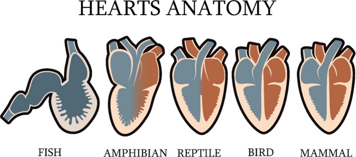
Convergence is the tendency of independent species to evolve similarly when subjected to the same environmental conditions. The primitive blueprint for the circulatory system emerged around 700-600 Mya and exhibits diverse physiological adaptations across the radiations of vertebrates (Subphylum Vertebrata, Phylum Chordata). It has evolved from the early chordate circulatory system with a single layered tube in the tunicate (Subphylum Urchordata) or an amphioxus (Subphylum Cephalochordata), to a vertebrate circulatory system with a two-chambered heart made up of one atrium and one ventricle in gnathostome fish (Infraphylum Gnathostomata), to a system with a three-chambered heart made up of two atria which maybe partially divided or completely separated in amphibian tetrapods (Class Amphibia). Subsequent tetrapods, including crocodiles and alligators (Order Crocodylia, Subclass Crocodylomorpha, Class Reptilia), birds (Subclass Aves, Class Reptilia) and mammals (Class Mammalia) evolved a four-chambered heart. The structure and function of the circulatory system of each individual holds a vital role which benefits each species specifically. The special characteristics of the four-chamber mammalian heart are highlighted by the peculiar structure of the myocardial muscle.
Keywords: circulatory system; comparative anatomy; evolution; heart; vertebrate.
© 2017 Anatomical Society.
Figures
Comment in
-
Evolution of the vertebrate heart.J Anat. 2018 May;232(5):886-887. doi: 10.1111/joa.12790. Epub 2018 Feb 27. J Anat. 2018. PMID: 29488213 Free PMC article. No abstract available.
-
Rebuttal letter in response to Professor R.H. Anderson's letter 'Evolution of the vertebrate heart'.J Anat. 2018 May;232(5):888-889. doi: 10.1111/joa.12788. Epub 2018 Feb 27. J Anat. 2018. PMID: 29488220 Free PMC article. No abstract available.
References
-
- Alexander RM. (ed.) 1986) The Collins Encyclopedia of Animal Biology. London: Collins.
-
- Anderson RH, Ho SY, Redmann K, et al. (2005) The anatomical arrangement of the myocardial cells making up the ventricular mass. Eur J Cardiothorac Surg 28, 517–525. - PubMed
-
- Axelsson M, Franklin CE (1997) From anatomy to angioscopy: 164 Years of crocodilian cardiovascular research, recent advances, and speculations. Comp Biochem Physiol A Physiol 118, 51–62.
-
- Bajaj P, Tang X, Saif TA, et al. (2010) Stiffness of the substrate influences the phenotype of embryonic chicken cardiac myocytes. J Biomed Mater Res A 95, 1261–1269. - PubMed
-
- Bardack D (1991) First fossil hagfish (myxinoidea), a record from the Pennsylvanian of Illinois. Science 254, 701–703. - PubMed
Publication types
MeSH terms
LinkOut - more resources
Full Text Sources
Other Literature Sources
