. 2017 Sep 14;12(9):e0183741.
doi: 10.1371/journal.pone.0183741. eCollection 2017.

Acute and chronic effects of treatment with mesenchymal stromal cells on LPS-induced pulmonary inflammation, emphysema and atherosclerosis development

Affiliations

Acute and chronic effects of treatment with mesenchymal stromal cells on LPS-induced pulmonary inflammation, emphysema and atherosclerosis development

P Padmini S J Khedoe et al. PLoS One. .

Abstract

Background: COPD is a pulmonary disorder often accompanied by cardiovascular disease (CVD), and current treatment of this comorbidity is suboptimal. Systemic inflammation in COPD triggered by smoke and microbial exposure is suggested to link COPD and CVD. Mesenchymal stromal cells (MSC) possess anti-inflammatory capacities and MSC treatment is considered an attractive treatment option for various chronic inflammatory diseases. Therefore, we investigated the immunomodulatory properties of MSC in an acute and chronic model of lipopolysaccharide (LPS)-induced inflammation, emphysema and atherosclerosis development in APOE*3-Leiden (E3L) mice.

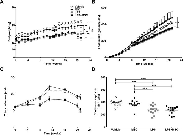
Methods: Hyperlipidemic E3L mice were intranasally instilled with 10 μg LPS or vehicle twice in an acute 4-day study, or twice weekly during 20 weeks Western-type diet feeding in a chronic study. Mice received 0.5x10⁶ MSC or vehicle intravenously twice after the first LPS instillation (acute study) or in week 14, 16, 18 and 20 (chronic study). Inflammatory parameters were measured in bronchoalveolar lavage (BAL) and lung tissue. Emphysema, pulmonary inflammation and atherosclerosis were assessed in the chronic study.

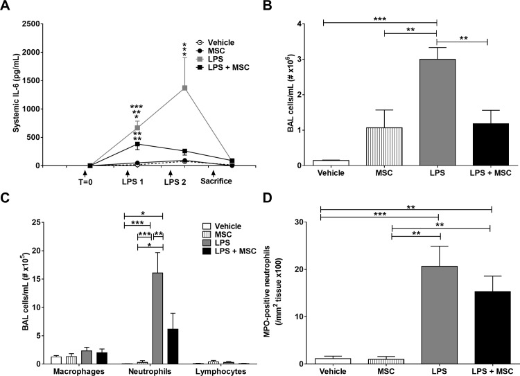
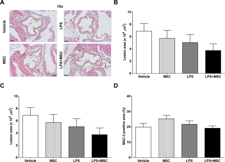
Results: In the acute study, intranasal LPS administration induced a marked systemic IL-6 response on day 3, which was inhibited after MSC treatment. Furthermore, MSC treatment reduced LPS-induced total cell count in BAL due to reduced neutrophil numbers. In the chronic study, LPS increased emphysema but did not aggravate atherosclerosis. Emphysema and atherosclerosis development were unaffected after MSC treatment.

Conclusion: These data show that MSC inhibit LPS-induced pulmonary and systemic inflammation in the acute study, whereas MSC treatment had no effect on inflammation, emphysema and atherosclerosis development in the chronic study.


Conflict of interest statement

Competing Interests: The authors have declared that no competing interests exist.

Figures

Fig 1
Fig 1. MSC treatment reduces inflammation in acute LPS-induced pulmonary inflammation.
APOE*3-Leiden (E3L) mice fed a Western-type diet (WTD) were intranasally instilled with vehicle or 10 μg LPS on day 1 and 3. Mice received 0.5x10⁶ MSC 4 h and 24 h after the first LPS instillation. Blood was drawn 3 h after LPS instillation to measure systemic IL-6 response (A). Mice were sacrificed on day 4 after which BAL was collected and total (B) and differential (C) cell number was counted. The number of MPO-positive neutrophils in the lungs was determined immunohistochemically (D). Data are shown as mean±SEM; n = 12–15; *p<0.05, **p<0.01, ***p<0.001.
Fig 2
Fig 2. MSC treatment does not affect chronic LPS-induced lowering in body weight, food intake and cholesterol.
E3L mice were intranasally instilled with vehicle or 10 μg LPS twice weekly during 20 weeks WTD feeding. Mice received vehicle or 0.5x10⁶ MSC in week 14, 16, 18 and 20. Bodyweight (A) and food intake (B) were monitored twice weekly during the study. Blood was drawn every 4 weeks to measure systemic cholesterol levels (C), which were used to calculate cholesterol exposure (D). Data are shown as mean±SEM; n = 12–15; *p<0.05, **p<0.01, ***p<0.001.
Fig 3
Fig 3. LPS exposure or MSC treatment do not affect blood leukocytes.
E3L mice were intranasally instilled with vehicle or 10 μg LPS twice weekly during 20 weeks WTD feeding. Mice received vehicle or 0.5x10⁶ MSC in week 14, 16, 18 and 20. Blood was drawn at 12 and 19 weeks and blood leukocytes, including WBC counts (A and B) were measured using an automated cell counter (Sysmex) and FACS analysis. (C) Levels of SAA were measured in plasma of mice at week 11 and week 16 (before and after MSC treatment). Data for A and B are shown as mean±SEM; n = 6–8; *p<0.05. Data in C are shown as mean±SEM; n = 12–15; ***p<0.001.
Fig 4
Fig 4. MSC treatment does not affect chronic LPS-induced decreased lung function.
E3L mice were intranasally instilled with vehicle or 10 μg LPS twice weekly during 20 weeks WTD feeding. Mice received vehicle or 0.5x10⁶ MSC in week 14, 16, 18 and 20. Total respiratory amplitude was measured at 12 and 20 weeks (A and B) using non-invasive whole body phlethysmography. Data are shown as mean±SEM; n = 12–15; *p<0.05, **p<0.01.
Fig 5
Fig 5. MSC treatment does not affect chronic LPS-induced emphysema, remodelling and inflammation.
After sacrifice, lungs were fixed in situ by gentle infusion of fixative by a continuous-release pump under constant pressure (through a tracheal cannula) after which paraffin sections were prepared and stained with HE (A). Emphysema was assessed by morphometric assessment of the MLI (B), as a measurement for destruction of alveolar walls and enlargement of alveolar space. In addition, remodelling (C) and inflammatory aggregates (D) were semi-quantified on Azan-trichrome stained lung slides. Data are shown as mean±SEM; n = 12–15; *p<0.05, **p<0.01, ***p<0.001.
Fig 6
Fig 6. MSC treatment does not affect chronic LPS-induced pulmonary inflammation.
E3L mice fed a WTD were intranasally instilled with vehicle or 10 μg LPS twice weekly during 20 weeks WTD feeding. Mice received vehicle or 0.5x10⁶ MSC in week 14, 16, 18 and 20. At sacrifice, BAL was collected for cytospin preparation and FACS analysis. Total cell number (A) and cell differentials (B) were obtained. BAL cells were also analysed using FACS analysis (C). At sacrifice, single cell suspensions were obtained from lung tissue and analysed by FACS (D). Data are shown as mean±SEM; n = 12–15;**p<0.01, ***p<0.001.
Fig 7
Fig 7. Chronic LPS instillation and MSC treatment do not affect atherosclerosis.
E3L mice fed a WTD were intranasally instilled with vehicle or 10 μg LPS twice weekly during 20 weeks WTD feeding. Mice received vehicle or 0.5x10⁶ MSC in week 14, 16, 18 and 20. Hearts were isolated and fixed in phosphate-buffered 4% formaldehyde and processed for paraffin embedding. For quantification and classification of atherosclerosis, the hearts were cross-sectioned (5 μm) throughout the entire aortic root area and stained with HPS (A). Lesion area (B) and severity (C) was determined with ImageJ. MAC-3 positive macrophages content was determined (D). Data are shown as mean±SEM; n = 12–15.

References

    1. From the Global Strategy for the Diagnosis, Management and Prevention of COPD, Global Initiative for Chronic Obstructive Lung Disease (GOLD) 2016. Available from: http://www.goldcopd.org/. 2016. - PubMed
    1. Vivodtzev I, Tamisier R, Baguet JP, Borel JC, Levy P, Pepin JL. Arterial stiffness in COPD. Chest. 2014;145(4):861–75. doi: 1852921 [pii]; doi: 10.1378/chest.13-1809 - DOI - PubMed
    1. Nussbaumer-Ochsner Y, Rabe KF. Systemic manifestations of COPD. Chest. 2011;139(1):165–73. doi: 139/1/165 [pii]; doi: 10.1378/chest.10-1252 - DOI - PubMed
    1. Libby P, Hansson GK. Inflammation and immunity in diseases of the arterial tree: players and layers. Circ Res. 2015;116(2):307–11. doi: CIRCRESAHA.116.301313 [pii]; doi: 10.1161/CIRCRESAHA.116.301313 - DOI - PMC - PubMed
    1. Barua RS, Sharma M, Dileepan KN. Cigarette Smoke Amplifies Inflammatory Response and Atherosclerosis Progression Through Activation of the H1R-TLR2/4-COX2 Axis. Front Immunol. 2015;6:572 doi: 10.3389/fimmu.2015.00572 ; PubMed Central PMCID: PMCPMC4638143. - DOI - PMC - PubMed

MeSH terms