

Role of the central lysine cluster and scrapie templating in the transmissibility of synthetic prion protein aggregates
- PMID: 28910420
- PMCID: PMC5614645
- DOI: 10.1371/journal.ppat.1006623
Role of the central lysine cluster and scrapie templating in the transmissibility of synthetic prion protein aggregates
Abstract
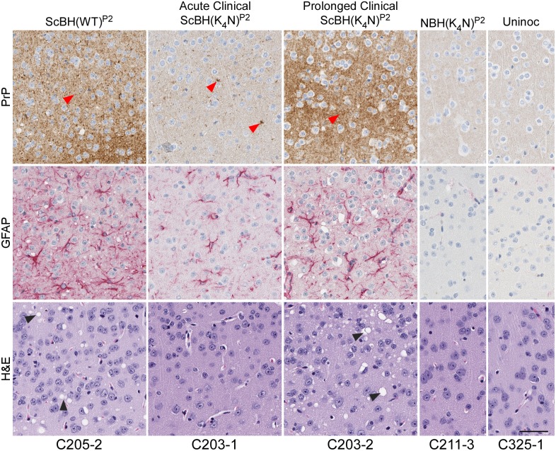
Mammalian prion structures and replication mechanisms are poorly understood. Most synthetic recombinant prion protein (rPrP) amyloids prepared without cofactors are non-infectious or much less infectious than bona fide tissue-derived PrPSc. This effect has been associated with differences in folding of the aggregates, manifested in part by reduced solvent exclusion and protease-resistance in rPrP amyloids, especially within residues ~90-160. Substitution of 4 lysines within residues 101-110 of rPrP (central lysine cluster) with alanines (K4A) or asparagines (K4N) allows formation of aggregates with extended proteinase K (PK) resistant cores reminiscent of PrPSc, particularly when seeded with PrPSc. Here we have compared the infectivity of rPrP aggregates made with K4N, K4A or wild-type (WT) rPrP, after seeding with scrapie brain homogenate (ScBH) or normal brain homogenate (NBH). None of these preparations caused clinical disease on first passage into rodents. However, the ScBH-seeded fibrils (only) led to a subclinical pathogenesis as indicated by increases in prion seeding activity, neuropathology, and abnormal PrP in the brain. Seeding activities usually accumulated to much higher levels in animals inoculated with ScBH-seeded fibrils made with the K4N, rather than WT, rPrP molecules. Brain homogenates from subclinical animals induced clinical disease on second passage into "hamsterized" Tg7 mice, with shorter incubation times in animals inoculated with ScBH-seeded K4N rPrP fibrils. On second passage from animals inoculated with ScBH-seeded WT fibrils, we detected an additional PK resistant PrP fragment that was similar to that of bona fide PrPSc. Together these data indicate that both the central lysine cluster and scrapie seeding of rPrP aggregates influence the induction of PrP misfolding, neuropathology and clinical manifestations upon passage in vivo. We confirm that some rPrP aggregates can initiate further aggregation without typical pathogenesis in vivo. We also provide evidence that there is little, if any, biohazard associated with routine RT-QuIC assays.
Conflict of interest statement
The authors have declared that no competing interests exist.
Figures
Similar articles
-
Structural biology of ex vivo mammalian prions.J Biol Chem. 2022 Aug;298(8):102181. doi: 10.1016/j.jbc.2022.102181. Epub 2022 Jun 23. J Biol Chem. 2022. PMID: 35752366 Free PMC article. Review.
-
PrP P102L and Nearby Lysine Mutations Promote Spontaneous In Vitro Formation of Transmissible Prions.J Virol. 2017 Oct 13;91(21):e01276-17. doi: 10.1128/JVI.01276-17. Print 2017 Nov 1. J Virol. 2017. PMID: 28835493 Free PMC article.
-
Prion seeding activities of mouse scrapie strains with divergent PrPSc protease sensitivities and amyloid plaque content using RT-QuIC and eQuIC.PLoS One. 2012;7(11):e48969. doi: 10.1371/journal.pone.0048969. Epub 2012 Nov 5. PLoS One. 2012. PMID: 23139828 Free PMC article.
-
Distinct structures of scrapie prion protein (PrPSc)-seeded versus spontaneous recombinant prion protein fibrils revealed by hydrogen/deuterium exchange.J Biol Chem. 2009 Sep 4;284(36):24233-41. doi: 10.1074/jbc.M109.036558. Epub 2009 Jul 13. J Biol Chem. 2009. PMID: 19596861 Free PMC article.
-
Immunoaffinity purification and neutralization of scrapie prions.Prog Clin Biol Res. 1989;317:583-600. Prog Clin Biol Res. 1989. PMID: 2574871 Review.
Cited by
-
Structural biology of ex vivo mammalian prions.J Biol Chem. 2022 Aug;298(8):102181. doi: 10.1016/j.jbc.2022.102181. Epub 2022 Jun 23. J Biol Chem. 2022. PMID: 35752366 Free PMC article. Review.
-
Challenges and Advances in Antemortem Diagnosis of Human Transmissible Spongiform Encephalopathies.Front Bioeng Biotechnol. 2020 Oct 20;8:585896. doi: 10.3389/fbioe.2020.585896. eCollection 2020. Front Bioeng Biotechnol. 2020. PMID: 33195151 Free PMC article. Review.
-
Preserving prion strain identity upon replication of prions in vitro using recombinant prion protein.Acta Neuropathol Commun. 2018 Sep 12;6(1):92. doi: 10.1186/s40478-018-0597-y. Acta Neuropathol Commun. 2018. PMID: 30208966 Free PMC article.
-
The prion 2018 round tables (I): the structure of PrPSc.Prion. 2019 Jan;13(1):46-52. doi: 10.1080/19336896.2019.1569450. Prion. 2019. PMID: 30646817 Free PMC article.
-
Cellulose ether treatment in vivo generates chronic wasting disease prions with reduced protease resistance and delayed disease progression.J Neurochem. 2020 Mar;152(6):727-740. doi: 10.1111/jnc.14877. Epub 2019 Oct 16. J Neurochem. 2020. PMID: 31553058 Free PMC article.
References
-
- Caughey BW, Dong A, Bhat KS, Ernst D, Hayes SF, Caughey WS. Secondary structure analysis of the scrapie-associated protein PrP 27–30 in water by infrared spectroscopy. Biochemistry. 1991;30:7672–80. - PubMed
-
- Bessen RA, Kocisko DA, Raymond GJ, Nandan S, Lansbury PT Jr., Caughey B. Nongenetic propagation of strain-specific phenotypes of scrapie prion protein. Nature. 1995;375:698–700. doi: 10.1038/375698a0 - DOI - PubMed
-
- Kocisko DA, Come JH, Priola SA, Chesebro B, Raymond GJ, Lansbury PT, et al. Cell-free formation of protease-resistant prion protein. Nature. 1994;370:471–4. doi: 10.1038/370471a0 - DOI - PubMed
-
- Telling GC, Parchi P, DeArmond SJ, Cortelli P, Montagna P, Gabizon R, et al. Evidence for the conformation of the pathologic isoform of the prion protein enciphering and propagating prion diversity. Science. 1996;274:2079–82. - PubMed
MeSH terms
Substances
LinkOut - more resources
Full Text Sources
Other Literature Sources
Molecular Biology Databases
Research Materials
