

Optical Coherence Tomography Angiography Features of Iris Racemose Hemangioma in 4 Cases
- PMID: 28910426
- PMCID: PMC5847105
- DOI: 10.1001/jamaophthalmol.2017.3390
Optical Coherence Tomography Angiography Features of Iris Racemose Hemangioma in 4 Cases
Abstract
Importance: Optical coherence tomography angiography (OCTA) allows visualization of iris racemose hemangioma course and its relation to the normal iris microvasculature.
Objective: To describe OCTA features of iris racemose hemangioma.
Design, setting, and participants: Descriptive, noncomparative case series at a tertiary referral center (Ocular Oncology Service of Wills Eye Hospital). Patients diagnosed with unilateral iris racemose hemangioma were included in the study.
Main outcomes and measures: Features of iris racemose hemangioma on OCTA.
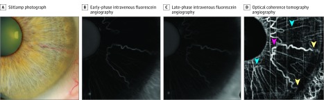
Results: Four eyes of 4 patients with unilateral iris racemose hemangioma were included in the study. Mean patient age was 50 years, all patients were white, and Snellen visual acuity was 20/20 in each case. All eyes had sectoral iris racemose hemangioma without associated iris or ciliary body solid tumor on clinical examination and ultrasound biomicroscopy. By anterior segment OCT, the racemose hemangioma was partially visualized in all cases. By OCTA, the hemangioma was clearly visualized as a uniform large-caliber vascular tortuous loop with intense flow characteristics superimposed over small-caliber radial iris vessels against a background of low-signal iris stroma. The vascular course on OCTA resembled a light bulb filament (filament sign), arising from the peripheral iris (base of light bulb) and forming a tortuous loop on reaching its peak (midfilament) near the pupil (n = 3) or midzonal iris (n = 1), before returning to the peripheral iris (base of light bulb). Intravenous fluorescein angiography performed in 1 eye depicted the iris hemangioma; however, small-caliber radial iris vessels were more distinct on OCTA than intravenous fluorescein angiography.
Conclusions and relevance: Optical coherence tomography angiography is a noninvasive vascular imaging modality that clearly depicts the looping course of iris racemose hemangioma. Optical coherence tomography angiography depicted fine details of radial iris vessels, not distinct on intravenous fluorescein angiography.
Conflict of interest statement
Figures

References
-
- Shields CL, Kancherla S, Patel J, et al. . Clinical survey of 3680 iris tumors based on patient age at presentation. Ophthalmology. 2012;119(2):407-414. - PubMed
-
- Shields JA, Streicher TF, Spirkova JH, Stubna M, Shields CL. Arteriovenous malformation of the iris in 14 cases. Arch Ophthalmol. 2006;124(3):370-375. - PubMed
-
- Lee BJ, Jeng BH, Singh AD. OCT and ultrasound biomicroscopic findings in iris arteriovenous malformation. Ophthalmic Surg Lasers Imaging. 2008;39(5):426-428. - PubMed
-
- Ang M, Sim DA, Keane PA, et al. . Optical coherence tomography angiography for anterior segment vasculature imaging. Ophthalmology. 2015;122(9):1740-1747. - PubMed
-
- Ang M, Cai Y, MacPhee B, et al. . Optical coherence tomography angiography and indocyanine green angiography for corneal vascularisation. Br J Ophthalmol. 2016;100(11):1557-1563. - PubMed
Publication types
MeSH terms
LinkOut - more resources
Full Text Sources
Other Literature Sources
Medical
