

Spectral-Domain Optical Coherence Tomographic Angiography in Children With Amblyopia
- PMID: 28910439
- PMCID: PMC5710487
- DOI: 10.1001/jamaophthalmol.2017.3423
Spectral-Domain Optical Coherence Tomographic Angiography in Children With Amblyopia
Abstract
Importance: Amblyopia is the most common cause of visual impairment in childhood, with a prevalence of 1% to 4% in children in the United States. To date, no studies using noninvasive optical coherence tomographic angiography (OCTA) have measured blood flow in the retinal capillary layers in children with amblyopia.
Objective: To evaluate the retinal and microvascular features using OCTA in children (<18 years) with amblyopia.
Design, setting, and participants: This observational case-control study enrolled patients from September 1, 2016, through May 31, 2017, and was conducted from September 1, 2016, through June 30, 2017, at the Stein Eye Institute at UCLA (University of California, Los Angeles). Participants included 59 children (<18 years) with amblyopia and without amblyopia examined at a pediatric ophthalmology clinic or referred to the clinic by coinvestigators. All patients underwent comprehensive ophthalmological examination, including visual acuity, refraction, and ocular motility tests; anterior and posterior segment examination; and OCTA.
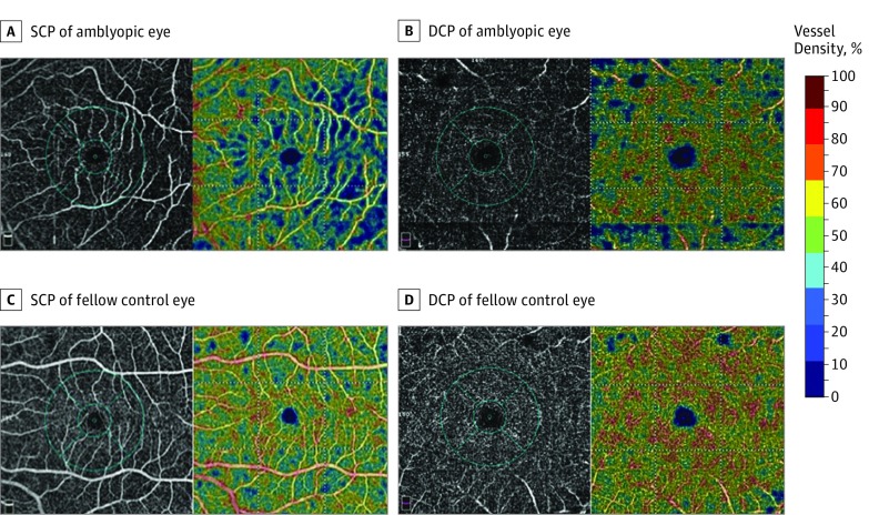
Main outcomes and measures: Reduced superficial and deep retinal capillary vessel density on OCTA.
Results: Of the 63 eyes evaluated, 13 (21%) were amblyopic and 50 (79%) were control eyes. Of the 59 patients, the mean (SD) age of patients with amblyopia was 8.0 (4.0) years and 10.3 (3.3) years for the controls; 33 patients (56%) were female; and 5 of 13 (39%) and 27 of 46 (54%) patients in the amblyopic and control groups, respectively, were identified as white. The macular vessel density of the superficial capillary plexus was lower in the amblyopic group than in the control group in both 3 × 3-mm and 6 × 6-mm scans. After adjusting for age and refractive error, the mean (SD) difference in the superficial capillary plexus in the 6 × 6-mm scan was statistically significant (49.3% [4.1] vs 51.2% [2.9]; P = .02). Macular vessel density of the deep capillary plexus in the 6 × 6-mm scans was also considerably different between groups: mean (SD) vessel density of the deep retinal capillary plexus was 54.4% (4.7%) in the amblyopia group and 60.1% (3.3%) in the control group, with a difference of 5.7% (95% CI, 3.4%-8.1%; P = .002).
Conclusions and relevance: The study found that OCTA reveals subnormal superficial and deep retinal capillary density in the macula of patients with amblyopia. Further studies are needed to determine the clinical relevance of this finding.
Conflict of interest statement
Figures

Similar articles
-
Foveal avascular zone and macular vessel density after correction for magnification error in unilateral amblyopia using optical coherence tomography angiography.BMC Ophthalmol. 2019 Aug 5;19(1):171. doi: 10.1186/s12886-019-1177-z. BMC Ophthalmol. 2019. PMID: 31382925 Free PMC article.
-
Association of Optical Coherence Tomography Angiography Metrics With Detection of Impaired Macular Microvasculature and Decreased Vision in Amblyopic Eyes: The Hong Kong Children Eye Study.JAMA Ophthalmol. 2020 Aug 1;138(8):858-865. doi: 10.1001/jamaophthalmol.2020.2220. JAMA Ophthalmol. 2020. PMID: 32584368 Free PMC article.
-
Comparison of Retinal Vascular Structure in Eyes With and Without Amblyopia by Optical Coherence Tomography Angiography.J Pediatr Ophthalmol Strabismus. 2020 Jan 1;57(1):48-53. doi: 10.3928/01913913-20191004-01. J Pediatr Ophthalmol Strabismus. 2020. PMID: 31972041
-
Evaluation of Retinal Microvascular Features in Patients with Amblyopia Based on Optical Coherence Tomography Angiography: A Systematic Review and Meta-Analysis.Ophthalmic Res. 2023;66(1):862-877. doi: 10.1159/000529857. Epub 2023 Mar 14. Ophthalmic Res. 2023. PMID: 36917963
-
Analysis of Retinal Microvasculature Features in Amblyopic Eyes: A Meta-Analysis.Ophthalmic Res. 2023;66(1):131-143. doi: 10.1159/000526531. Epub 2022 Aug 23. Ophthalmic Res. 2023. PMID: 35998587
Cited by
-
Microvasculature evaluation of anisometropic amblyopia children by Angio-OCT.Sci Rep. 2023 Feb 16;13(1):2780. doi: 10.1038/s41598-023-29816-1. Sci Rep. 2023. PMID: 36797301 Free PMC article.
-
Ultra-Widefield Swept-Source Optical Coherence Tomography Angiography in the Assessment of Choroidal Changes in Young Adults With Myopia.Transl Vis Sci Technol. 2022 Dec 1;11(12):14. doi: 10.1167/tvst.11.12.14. Transl Vis Sci Technol. 2022. PMID: 36580322 Free PMC article.
-
OCT-Angiography Findings in Patients with Amblyopia: Comparison between Healthy Controls, Treatment-Responsive, and Treatment-Unresponsive Amblyopic Patients.Diagnostics (Basel). 2021 Sep 24;11(10):1751. doi: 10.3390/diagnostics11101751. Diagnostics (Basel). 2021. PMID: 34679448 Free PMC article.
-
Optical Coherence Tomography and Optical Coherence Tomography Angiography in Pediatric Retinal Diseases.Diagnostics (Basel). 2023 Apr 18;13(8):1461. doi: 10.3390/diagnostics13081461. Diagnostics (Basel). 2023. PMID: 37189561 Free PMC article. Review.
-
Acupuncture for pediatric bilateral amblyopia.Integr Med Res. 2020 Dec;9(4):100435. doi: 10.1016/j.imr.2020.100435. Epub 2020 May 30. Integr Med Res. 2020. PMID: 32760651 Free PMC article. No abstract available.
References
-
- Repka M. Amblyopia: basics, questions, and practical management In: Lyons C, Lambert S, eds. Taylor and Hoyt’s Pediatric Ophthalmology and Strabismus. 5th ed Amsterdam, Netherlands: Elsevier; 2017:754-761.
-
- Williams C, Northstone K, Howard M, Harvey I, Harrad RA, Sparrow JM. Prevalence and risk factors for common vision problems in children: data from the ALSPAC study. Br J Ophthalmol. 2008;92(7):959-964. - PubMed
-
- Li J, Ji P, Yu M. Meta-analysis of retinal changes in unilateral amblyopia using optical coherence tomography. Eur J Ophthalmol. 2015;25(5):400-409. - PubMed
-
- Gunton KB. Advances in amblyopia: what have we learned from PEDIG trials? Pediatrics. 2013;131(3):540-547. - PubMed
Publication types
MeSH terms
LinkOut - more resources
Full Text Sources
Other Literature Sources
Medical
