Review
. 2017 Sep 13;95(6):1246-1265.
doi: 10.1016/j.neuron.2017.07.010.

Neuroimmunology of Traumatic Brain Injury: Time for a Paradigm Shift

Affiliations
Review

Neuroimmunology of Traumatic Brain Injury: Time for a Paradigm Shift

Yasir N Jassam et al. Neuron. .

Abstract

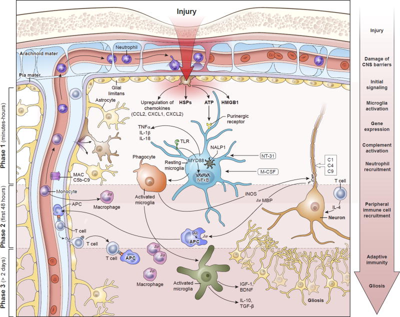
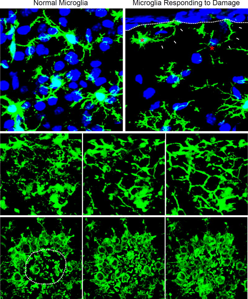
Traumatic brain injury (TBI) is a leading cause of morbidity and disability, with a considerable socioeconomic burden. Heterogeneity of pathoanatomical subtypes and diversity in the pathogenesis and extent of injury contribute to differences in the course and outcome of TBI. Following the primary injury, extensive and lasting damage is sustained through a complex cascade of events referred to as "secondary injury." Neuroinflammation is proposed as an important manipulable aspect of secondary injury in animal and human studies. Because neuroinflammation can be detrimental or beneficial, before developing immunomodulatory therapies, it is necessary to better understand the timing and complexity of the immune responses that follow TBI. With a rapidly increasing body of literature, there is a need for a clear summary of TBI neuroimmunology. This review presents our current understanding of the immune response to TBI in a chronological and compartment-based manner, highlighting early changes in gene expression and initial signaling pathways that lead to activation of innate and adaptive immunity. Based on recent advances in our understanding of innate immune cell activation, we propose a new paradigm to study innate immune cells following TBI that moves away from the existing M1/M2 classification of activation states toward a stimulus- and disease-specific understanding of polarization state based on transcriptomic and proteomic profiling.

Keywords: M1; M2; TBI; astrocytes; microglia; models; neurodegeneration; neuroimmunology; neuroinflammation; neutrophils; transcriptome.


Figures

Figure 1
Figure 1. Temporal progression of the immune response to contusion TBI
Phase I begins within minutes of brain injury due to the release of alarmins from the damaged meninges, glial limitans, and parenchyma, such as ATP, HSPs, HGMB1, etc. These signals bind to PAMP and DAMP sensors like TLRs and purinergic receptors that induce immediate activation of resident myeloid cells (e.g. microglia) and inflammasome assembly (NALP1) that promotes the generation of mature IL-1β and IL-18. In addition, NFκB translocates to the nuclei of these cells and induces an immunological program involving cellular proliferation and the release inflammatory amplifiers such as chemokines, cytokines, ROS, and NO, among others. Phase 1 also includes complement activation and the recruitment of neutrophils to the meninges and perivascular spaces. Neutrophil recruitment depends in part on purinergic receptor signaling. Secondary damage to CNS tissue occurs in Phase 1 and can continue into Phase 2. This can be mediated by inflammatory cytokines, complement, and ROS. T cells and monocytes are recruited to the damage site in Phase 2, where monocytes convert into macrophages and T cells have the ability to produce neuroprotective cytokines in response to alarmins. Macrophages participate in the cleanup of debris and damaged cells. Based on their state of functional activation, they can either promote further damage or initiate the process of inflammatory resolution and tissue repair. Inflammation can continue for an extended period of time into Phase 3. Self-antigens released from damaged neural cells can be presented by local APCs to T cells. The ideal outcome during Phase 3 is resolution of the inflammatory response, release of trophic factors, and isolation damaged areas via astrocytes. However, this is does not always occur following TBI and chronic inflammation can persist. Abbreviations: APC, antigen-presenting cell; ATP, adenosine triphosphate; BDNF, brain-derived neurotrophic factor; CCL2, chemokine (C-C motif) ligand 2; CXCL1, chemokine (C-X-C motif) ligand 1; CXCL2, chemokine (C-X-C motif) ligand 2; DAMP, damage associated molecular pattern molecules; HSPs, heat shock proteins; HMGB1, high mobility group box 1 protein; IGF-1, insulin-like growth factor-1; IL-1, interleukin-1; IL-4, interleukin-4; IL-10, interleukin-10; iNOS, inducible nitric oxide synthase; MyD88, myeloid differentiation primary response gene 88; NALP1, NAcht leucine-rich repeat protein 1; NT-3, neurotrophin-3; NFκB, nuclear factor kappa-light-chain-enhancer of activated B cells; MBP, myelin basic protein; M-CSF, macrophage colony stimulating factor; NO, nitric oxide; PAMP, pathogen-associated molecular pattern; ROS, reactive oxygen species; TGF-β, transforming growth factor beta; TLR, toll-like receptors; TNF-α, tumor necrosis factor-alpha
Figure 2
Figure 2. Acute microglial dynamics following mild focal cortical injury
(A, B) Representative confocal images captured within the brain of a CX3CR1gfp/+ (green) mouse 3 hours following cortical injury show two microglia (red asterisks; panel B) extending processes (white arrowheads) toward the injured glial limitans (white dotted line) (Roth et al., 2014). Uninvolved, ramified microglia beneath the area of injury are shown in panel A for comparison. Cell nuclei are blue. (C, D) Representative time lapses captured by intravital two-photon microscopy through the thinned skull window of CX3CR1gfp/+ mice following mTBI. Panel C is a time lapse (beginning 5 min post-injury) depicting the morphological transformation of ramified microglia into “honeycomb” like structures that circumscribe individual astrocytes (white asterisks) within the injured glial limitans. These structures provide barrier support. Panel D shows the convergence of phagocytic “jellyfish” microglia into an area of heavy brain damage (white dotted line). These cells participate in the phagocytic clearance of debris.

References

    1. Adamczak S, Dale G, de Rivero Vaccari JP, Bullock MR, Dietrich WD, Keane RW. Inflammasome proteins in cerebrospinal fluid of brain-injured patients as biomarkers of functional outcome: clinical article. J Neurosurg. 2012;117:1119–1125. - PMC - PubMed
    1. Ahmad A, Crupi R, Campolo M, Genovese T, Esposito E, Cuzzocrea S. Absence of TLR4 reduces neurovascular unit and secondary inflammatory process after traumatic brain injury in mice. PLoS One. 2013;8:e57208. - PMC - PubMed
    1. Almeida-Suhett CP, Li Z, Marini AM, Braga MF, Eiden LE. Temporal course of changes in gene expression suggests a cytokine-related mechanism for long-term hippocampal alteration after controlled cortical impact. J Neurotrauma. 2014;31:683–690. - PMC - PubMed
    1. Ambrosini E, Aloisi F. Chemokines and glial cells: a complex network in the central nervous system. Neurochem Res. 2004;29:1017–1038. - PubMed
    1. Anthonymuthu TS, Kenny EM, Bayir H. Therapies targeting lipid peroxidation in traumatic brain injury. Brain Res. 2016;1640:57–76. - PMC - PubMed