. 2017 Sep 6;45(15):9138-9148.
doi: 10.1093/nar/gkx527.

Structural accommodation of ribonucleotide incorporation by the DNA repair enzyme polymerase Mu

Affiliations

Structural accommodation of ribonucleotide incorporation by the DNA repair enzyme polymerase Mu

Andrea F Moon et al. Nucleic Acids Res. .

Abstract

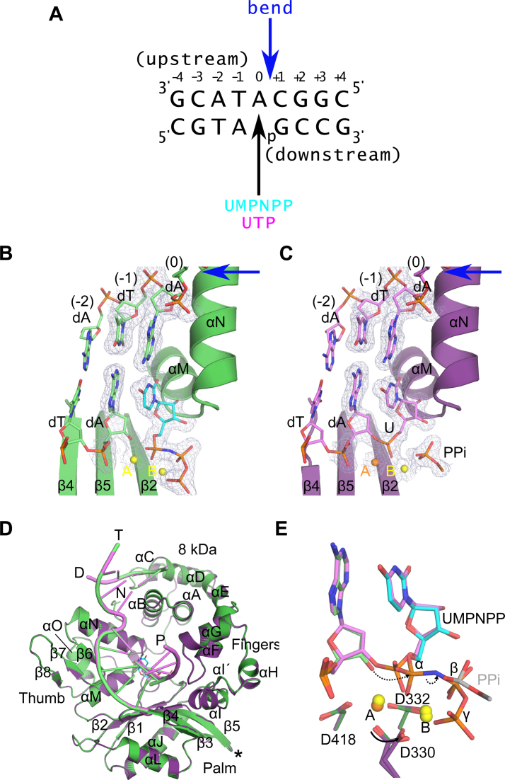
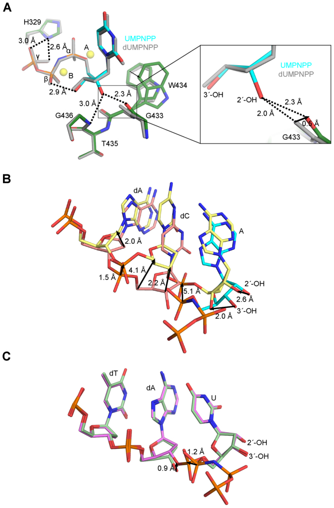
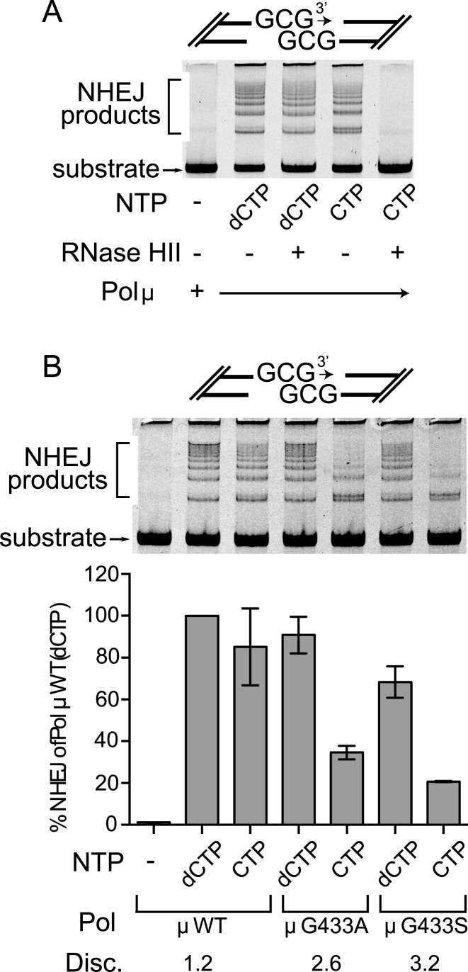
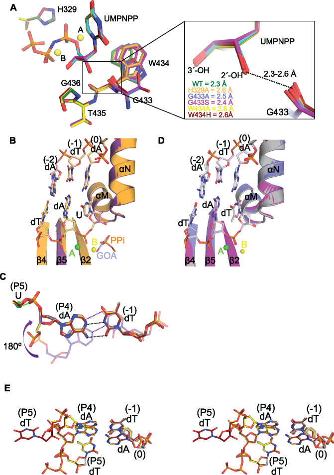
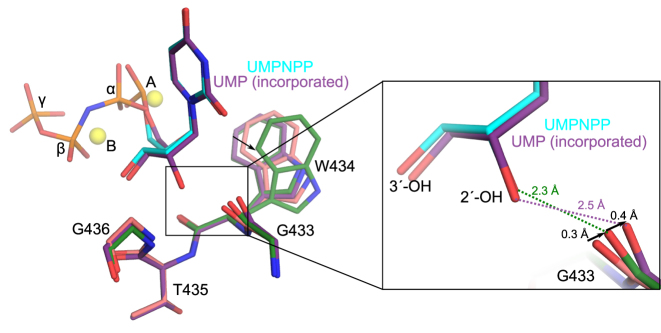
While most DNA polymerases discriminate against ribonucleotide triphosphate (rNTP) incorporation very effectively, the Family X member DNA polymerase μ (Pol μ) incorporates rNTPs almost as efficiently as deoxyribonucleotides. To gain insight into how this occurs, here we have used X-ray crystallography to describe the structures of pre- and post-catalytic complexes of Pol μ with a ribonucleotide bound at the active site. These structures reveal that Pol μ binds and incorporates a rNTP with normal active site geometry and no distortion of the DNA substrate or nucleotide. Moreover, a comparison of rNTP incorporation kinetics by wildtype and mutant Pol μ indicates that rNTP accommodation involves synergistic interactions with multiple active site residues not found in polymerases with greater discrimination. Together, the results are consistent with the hypothesis that rNTP incorporation by Pol μ is advantageous in gap-filling synthesis during DNA double strand break repair by nonhomologous end joining, particularly in nonreplicating cells containing very low deoxyribonucleotide concentrations.


Figures

Figure 1.
Figure 1.
Structural evaluation of ribonucleotide incorporation by wildtype hPol μΔ2. Wildtype hPol μΔ2 was crystallized in complex with a 1-nt gapped DNA substrate and an incoming dUMPNPP (A). A pre-catalytic ternary complex (B, green) was obtained by soaking with nonhydrolyzable UMPNPP (cyan), and a post-catalytic nicked complex (C, purple) was obtained by soaking with hydrolyzable UTP. Structures of the active sites of each complex are shown, with the protein drawn in cartoon and the DNA substrate in stick. Protein secondary structural elements are marked, with β-strands numbered, and α-helices labeled alphabetically. Magnesium (yellow) and manganese (orange) ions are drawn as spheres. 2FoFc electron density (contoured at 1σ) is shown for the DNA substrate near the catalytic center of each complex. The location of the 90° bend in the DNA template strand is indicated by a blue arrow. The partially disordered pyrophosphate leaving group is shown in stick. (D) Global superpositions of the pre- (green) and post-catalytic (purple) complexes. The location of the Loop2 truncation is shown as a black asterisk. (E) Detailed comparison of the wildtype hPol μΔ2 active center in the pre- (protein in dark green; primer terminus in light green; UMPNPP in cyan), and post-catalytic (protein in purple; DNA in lavender) complexes. Dashed arrows indicate movement of electrons during progression of the reaction.  All structural figures were generated using PyMOL (Schrödinger).
Figure 2.
Figure 2.
Analysis of interactions involving the 2′-OH on the incoming ribonucleotide. (A) Superposition of pre-catalytic ternary complexes of wildtype hPol μΔ2 with incoming deoxy- (dUMPNPP, gray) or ribonucleotide (protein in dark green; UMPNPP in cyan). Magnesium ions (yellow) from the ribonucleotide bound structure are shown for orientation. Zoomed in view (right) of putative hydrogen bonding interactions (dashed lines) with the 2′-OH. (B) Superposition of the Pol λ Loop1 deletion variant pre- (pink, PDB ID code 3UPQ (33); AMPNPP in cyan) and post- catalytic (yellow, PDB ID code 3UQ0 (33)) complexes.  (C) Superposition of the hPol μΔ2 pre- (light green) and post-catalytic (lavender) complexes. Direction and calculated distances for movement of paired atoms are shown as black arrows.
Figure 3.
Figure 3.
Comparison of nucleotide discrimination by Pol μ in NHEJ. (A) In vitro NHEJ concatamerization assay for full-length wildtype Pol μ, using either dCTP or CTP, as indicated. Following completion of NHEJ, dCTP- or CTP-incorporated reaction mixtures were treated with RNase HII, which cleaves where ribonucleotides have been incorporated into DNA (52). (B) Comparison of nucleotide discrimination by wildtype or mutant Pol μ in the same in vitro NHEJ concatamerization assay used above (top), lacking only the RNase HII treatment step. Extent of end-joining by each protein variant was quantitated by real-time PCR (bottom). All values represent calculated averages of end-joining (normalized to the activity of wildtype Pol μ with dCTP incorporation) ± the standard deviation for each experiment (Supplementary Table S4). Each assay was performed in triplicate.
Figure 4.
Figure 4.
Structural characterization of Pol μ ribonucleotide discrimination mutants. (A) Structural superpositions of the active site residues in the pre-catalytic ternary complexes of hPol μΔ2 wildtype (green, UMPNPP in cyan) and ribonucleotide discrimination mutants (H329A in orange, G433A in blue, G433S in magenta, W434A in yellow, W434H in maroon). The two magnesium ions (yellow spheres) from the wildtype structure are shown for orientation. The panel on the right gives a magnified view of the 2′-OH distances to the carbonyl of residue 433. (B) Structural superpositions of the post-catalytic complexes of the hPol μΔ2 wildtype (protein in purple, DNA in lavender) and H329A mutant (protein in orange, DNA in khaki). The partially disordered pyrophosphate leaving group (PPi, orange) from the wildtype structure, and a bound glycolate molecule (GOA, gray) from the H329A structure are modeled in the active site in stick. (C) Stick diagram of the expulsion of the newly incorporated rUMP from the active sites of the G433A/S (G433A in blue, G433S in magenta) and W434A/H (W434A in yellow, W434H in maroon) mutants. The canonical position maintained by the wildtype protein (purple, transparent) is shown for comparison. Putative hydrogen bonding interactions for the wildtype (purple dashes) and mutant (black dashes) base pair are indicated. Coordination of the HhH2 sodium ion (green sphere) by the phosphate between residues P4 and P5 in the mutants is drawn as a green line. (D) Structural superpositions of the post-catalytic complexes of the hPol μΔ2 wildtype (gray), G433A (blue), and G433S (magenta) mutants with incorporated dTMP. E. Stereo diagram of DNA substrate mixtures from the post-catalytic complexes of the hPol μΔ2 wildtype (gray, PDB ID code 4M0A (35)), W434A (yellow), and W434H (maroon) mutants with incoming dTTP/incorporated dTMP.
Figure 5.
Figure 5.
Putative role of Gly433 backbone carbonyl motions for ribonucleotide binding and incorporation. Structural superposition of hPol μΔ2 1-nt gapped binary (pink, PDB ID code 1LZG (35)), pre-catalytic ternary (protein in green; UMPNPP in cyan), and post-catalytic nicked (purple) complexes. Magnesium ions (yellow) from the pre-catalytic structure are shown for orientation. Global view of regions in close proximity to the 2′-OH are shown on the left, with a zoomed-in view (right) of putative hydrogen bonding interactions (dashed lines) with the 2′-OH.

References

    1. Bebenek K., Kunkel T.A.. Functions of DNA polymerases. Adv. Protein Chem. 2004; 69:137–165. - PubMed
    1. Nick McElhinny S.A., Watts B.E., Kumar D., Watt D.L., Lundstrom E.B., Burgers P.M., Johansson E., Chabes A., Kunkel T.A.. Abundant ribonucleotide incorporation into DNA by yeast replicative polymerases. Proc. Natl. Acad. Sci. U.S.A. 2010; 107:4949–4954. - PMC - PubMed
    1. Clark A.B., Lujan S.A., Kissling G.E., Kunkel T.A.. Mismatch repair-independent tandem repeat sequence instability resulting from ribonucleotide incorporation by DNA polymerase epsilon. DNA Repair (Amst.). 2011; 10:476–482. - PMC - PubMed
    1. Clausen A.R., Zhang S., Burgers P.M., Lee M.Y., Kunkel T.A.. Ribonucleotide incorporation, proofreading and bypass by human DNA polymerase delta. DNA Repair (Amst.). 2013; 12:121–127. - PMC - PubMed
    1. Traut T.W. Physiological concentrations of purines and pyrimidines. Mol. Cell. Biochem. 1994; 140:1–22. - PubMed

MeSH terms