. 2017 Sep 6;45(15):8957-8967.
doi: 10.1093/nar/gkx581.

The SmAP2 RNA binding motif in the 3'UTR affects mRNA stability in the crenarchaeum Sulfolobus solfataricus

Affiliations

The SmAP2 RNA binding motif in the 3'UTR affects mRNA stability in the crenarchaeum Sulfolobus solfataricus

Birgit Märtens et al. Nucleic Acids Res. .

Abstract

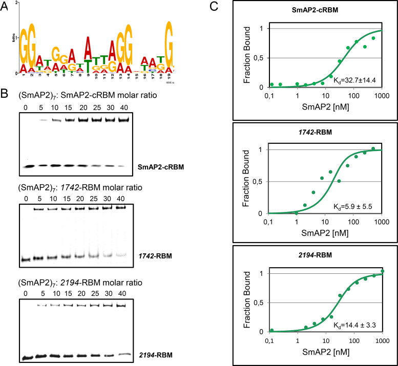
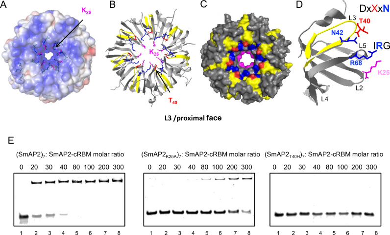
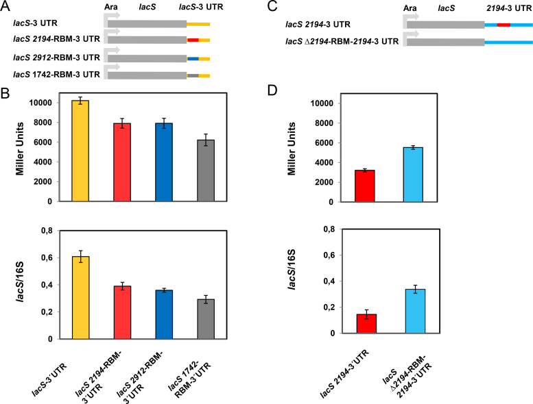
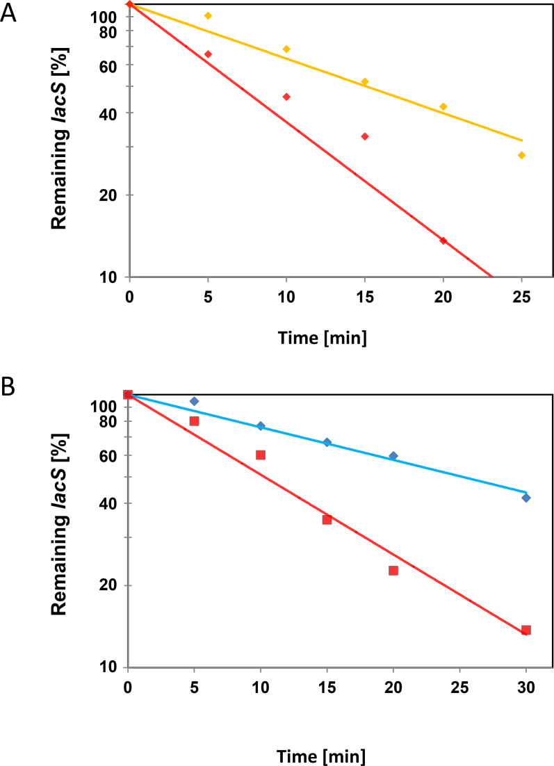
Sm and Sm-like proteins represent an evolutionarily conserved family with key roles in RNA metabolism in Pro- and Eukaryotes. In this study, a collection of 53 mRNAs that co-purified with Sulfolobus solfataricus (Sso) SmAP2 were surveyed for a specific RNA binding motif (RBM). SmAP2 was shown to bind with high affinity to the deduced consensus RNA binding motif (SmAP2-cRBM) in vitro. Residues in SmAP2 interacting with the SmAP2-cRBM were mapped by UV-induced crosslinking in combination with mass-spectrometry, and verified by mutational analyses. The RNA-binding site on SmAP2 includes a modified uracil binding pocket containing a unique threonine (T40) located on the L3 face and a second residue, K25, located in the pore. To study the function of the SmAP2-RBM in vivo, three authentic RBMs were inserted in the 3'UTR of a lacS reporter gene. The presence of the SmAP2-RBM in the reporter-constructs resulted in decreased LacS activity and reduced steady state levels of lacS mRNA. Moreover, the presence of the SmAP2-cRBM in and the replacement of the lacS 3'UTR with that of Sso2194 encompassing a SmAP2-RBM apparently impacted on the stability of the chimeric transcripts. These results are discussed in light of the function(s) of eukaryotic Lsm proteins in RNA turnover.


Figures

Figure 1.
Figure 1.
Binding of the SmAP2-RBM to SmAP2. (A) The consensus SmAP2 RNA binding motif (SmAP2-cRBM) was deduced from 53 mRNA sequences bound to SmAP2 (6) using the MEME online tool (28). (B) EMSA assays were performed with (SmAP2)7 and the RNA substrates SmAP2-cRBM (5′-GGAUGGAUAUUAGGAAAUG-3′; top panel), the 1742-RBM (5′-GGAGAUAUAGUAGGAUAG-3′; middle panel) and the 2194-RBM 5′-GGAUGGAUAUUAGGUACUGGG-3′; bottom panel). As described in Materials and Methods, increasing concentrations of (SmAP2)7 were incubated with the SmAP2-cRBM, the 1742-RBM and the 2194-RBM in a molar ratio of 0:1, 5:1, 10:1, 15:1, 20:1, 25:1, 30:1 and 40:1, respectively. (C) Dissociation constants (Kd's) of (SmAP2)7 for the SmAP2-cRBM (top panel), for the 1742-RBM (middle panel) and for the 2194-RBM (bottom panel) revealed by microscale thermophoresis (31). 25 nM fluorescently labelled SmAP2-cRBM (top panel), 1742-RBM (middle panel) and 2194-RBM (bottom panel) were added to increasing amounts of (SmAP2)7 protein. The Kd′s were determined as described in Materials and Methods, and were expressed as mean EC50 ± EC50 confidence interval of 2 independent experiments.
Figure 2.
Figure 2.
Location of residues in (SmAP2)7 and in the SmAP2 monomer interacting with uracil residue(s) of the SmAP2-cRBM revealed by UV-crosslinking and mass spectrometry. (A) Poisson–Boltzmann electrostatic potential of the solvent accessible L3/proximal face of (SmAP2)7 (15.) The location of K25 is highlighted in magenta. (B) Ribbon model of (SmAP2)7 depicting the location of K25 (magenta) and the peptide L34–R46 (yellow) involved in SmAP2-cRBM binding. The location of the T40 residue is indicated in red. (C) Space filling model of (SmAP2)7 depicting the locations of K25 (magenta), the peptide L34–R46 (yellow) and T40 (red). The SmAP2-specific T40 residue is located at the rim of the pore in the DxXxN motif, which has been described for eukaryal Lsm-complexes (10). N42 (blue) in the conserved DxXxN motif and R68 (blue) in the conserved IRG motif are involved in RNA binding in other Sm-proteins (15,22). (D) Location of residues involved in RNA-binding depicted in the SmAP2 monomer. The residues T40 (red) and N42 (blue) are located in the L3 loop in the conserved DxXxN motif, R68 (blue) is located in the L5 loop in the IRG motif and K25 (magenta) is located in the L2 domain. (E) EMSA assays performed with the (SmAP2K25A)7 and (SmAP2T40H)7 mutant proteins and the SmAP2-cRBM. Increasing concentrations of (SmAP2)7, (SmAP2K25A)7 and (SmAP2T40H)7, respectively were incubated with the SmAP2-cRBM in a molar ratio of 0:1 (lane1), 20:1 (lane 2), 30:1 (lane 3), 40:1 (lane 4), 80:1 (lane 5), 100:1 (lane 6), 200:1(lane 7) and 300:1 (lane 8), respectively.
Figure 3.
Figure 3.
Regulatory function of the SmAP2-cRBM motif. (A) Schematic depiction of the SSV-1 derived Sso-shuttle plasmid pMJ05, in which the lacS gene containing its authentic 3′UTR is preceded by an arabinose-inducible promoter (Ara). In the lacS 2194-RBM-3′UTR construct, the 2194-RBM (red line) identified in the 3′ UTR of the putative thermopsine gene (Sso2194) was transplanted to the 3′UTR of the lacS gene, 14 nt downstream of the G of the TAG stop codon. The 2912-RBM identified in the 3′ UTR of a sulfate adenylyltransferase gene (Sso2912) (blue line) and the 1742-RBM identified in the 3′ UTR of the terminal quinol oxidase gene (Sso1742) (grey line) were inserted analogously. (B) β-galactosidase activities (top panel) and lacS mRNA steady state levels (bottom panel) obtained with the Sso strain PH1–16 harbouring the constructs containing the lacS 3′UTR (pMJ05-Ara-lacS-3′UTR; orange bars), the 2194-RBM inserted in the lacS 3′UTR (pMJ05-Ara-lacS-2194-RBM-3′UTR; red bars), the 2912-RBM inserted in the lacS 3′UTR (pMJ05-Ara-lacS-2912-RBM-3′UTR; blue bars), and the 1742-RBM inserted in the lacS 3′UTR (pMJ05-Ara-lacS-1742-RBM-3′UTR; gray bars) respectively. The β-galactosidase activities and the lacS mRNA levels were determined as described in Materials and Methods. Each experiment was performed with three biological replicates of the respective strains. The error bars are derived from biological and technical replicates. The statistical significance was determined using a t-test (Supplementary Table S2). (C) Schematic depiction of the SSV-1 derived Sso-shuttle plasmid pMJ05, in which the lacS 3′UTR was replaced by the 2194–3′UTR (lacS 2194–3′UTR). In the lacS Δ2194-RBM-2194–3′UTR construct, the 2194-RBM 5′-GGAGATATAGTAGGTATAGTA-3′ was replaced by the unrelated sequence 5′-TTCTGGCCCACCATACCATCGC-3′. (D) β-galactosidase activities (top panel) and lacS mRNA steady state levels (bottom panel) obtained with the Sso strain PH1–16 harbouring the constructs containing the 2194–3′UTR abutted to the lacS gene (pMJ05-Ara-lacS-2194–3′UTR; red bars) and the Δ2194-RBM-2194–3′UTR (pMJ05-Ara-Δ2194-RBM-2194–3′UTR; light blue bars), respectively. The ß-galactosidase activities and the lacS mRNA levels were determined as described in Materials and Methods. Each experiment was performed with three biological replicates of the respective strain. The error bars are derived from biological and technical replicates. The statistical significance was determined using a t-test (Supplementary Table S2).
Figure 4.
Figure 4.
Reduced lacS mRNA levels upon placement of the 2194-RBM in its 3′UTR. (A) The Sso strains PH1–16(pMJ05-Ara-lacS-3′UTR) and PH1–16(pMJ05-Ara-lacS-2194-RBM-3′UTR) were grown in Brock's medium at 75°C containing 0.2% arabinose to an OD600 of 0.5. Then, the cells were harvested and resuspended in the same volume of Brock's medium containing 0.2% sucrose pre-warmed to 75°C ( = 0 min). After 5, 10, 15, 20 and 25 min, total RNA was isolated and cDNA was generated as described in Materials and Methods. The remaining lacS mRNA levels after promoter shut-off were quantified by qPCR using cDNA derived from Sso PH1–16 harboring either plasmid pMJ05-Ara-lacS-3′UTR (orange graph) or pMJ05-Ara-lacS-2194-RBM-3′UTR (red graph) and normalized to 16S rRNA levels. The experiment was performed with two biological and three technical replicates. A least square analysis of semilogarithmic plots of remaining lacS mRNA versus time is shown. The respective lacS mRNA levels at time 0 were set to 100%. (B) The experiment was performed as described in A, except that the strains PH1–16(pMJ05-Ara-lacS-2194–3′UTR) (red graph) and PH1–16(pMJ05-Ara-Δ2194-RBM-2194–3′UTR) (light blue graph) were used and a 30 min time point was included.

Similar articles

Cited by

References

    1. Wilusz C.J., Wilusz J.. Lsm proteins and Hfq: Life at the 3′ end. RNA Biol. 2013; 10:592–601. - PMC - PubMed
    1. Collins B.M., Harrop S.J., Kornfeld G.D., Dawes I.W., Curmi P.M., Mabbutt B.C.. Crystal structure of a heptameric Sm-like protein complex from archaea: implications for the structure and evolution of snRNPs. J. Mol. Biol. 2001; 309:915–923. - PubMed
    1. Mura C., Cascio D., Sawaya M.R., Eisenberg D.S.. The crystal structure of a heptameric archaeal Sm protein: implications for the eukaryotic snRNP core. Proc. Natl. Acad. Sci. U.S.A. 2001; 98:5532–5537. - PMC - PubMed
    1. Törö I., Thore S., Mayer C., Basquin J., Seraphin B., Suck D.. RNA binding in an Sm core domain: X-ray structure and functional analysis of an archaeal Sm protein complex. EMBO J. 2001; 20:2293–2303. - PMC - PubMed
    1. Törö I., Basquin J., Teo-Dreher H., Suck D.. Archaeal Sm proteins form heptameric and hexameric complexes: crystal structures of the Sm1 and Sm2 proteins from the hyperthermophile Archaeoglobus fulgidus. J. Mol. Biol. 2002; 320:129–142. - PubMed

MeSH terms