. 2017 Sep 6;45(15):8993-9004.
doi: 10.1093/nar/gkx622.

The IRES5'UTR of the dicistrovirus cricket paralysis virus is a type III IRES containing an essential pseudoknot structure

Affiliations

The IRES5'UTR of the dicistrovirus cricket paralysis virus is a type III IRES containing an essential pseudoknot structure

Lauriane Gross et al. Nucleic Acids Res. .

Abstract

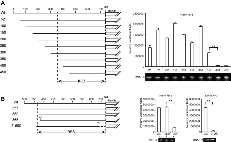
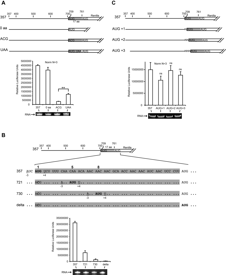
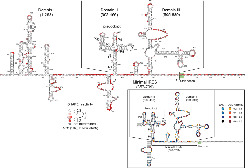
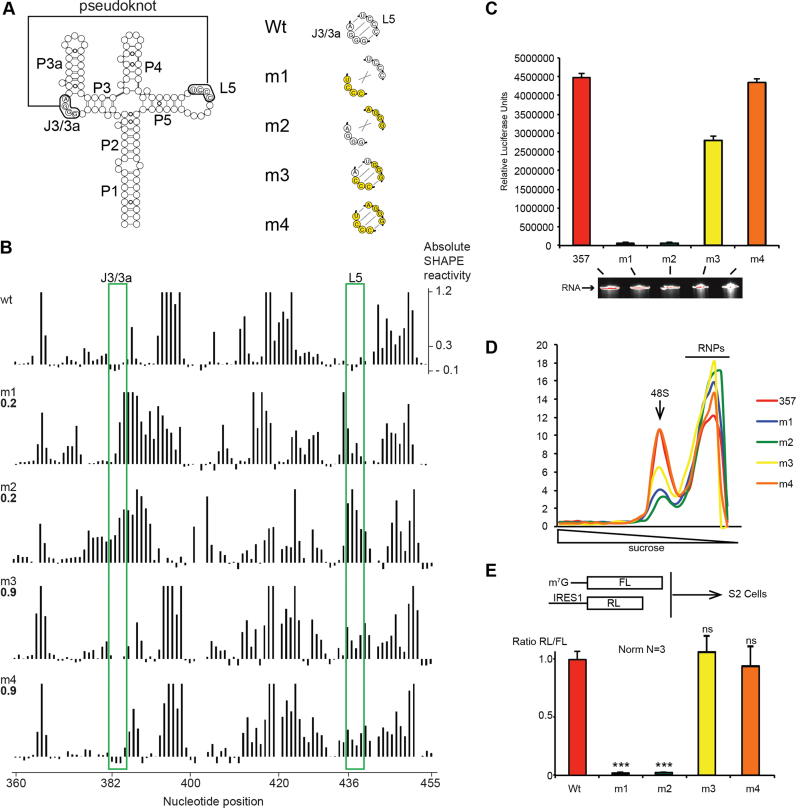
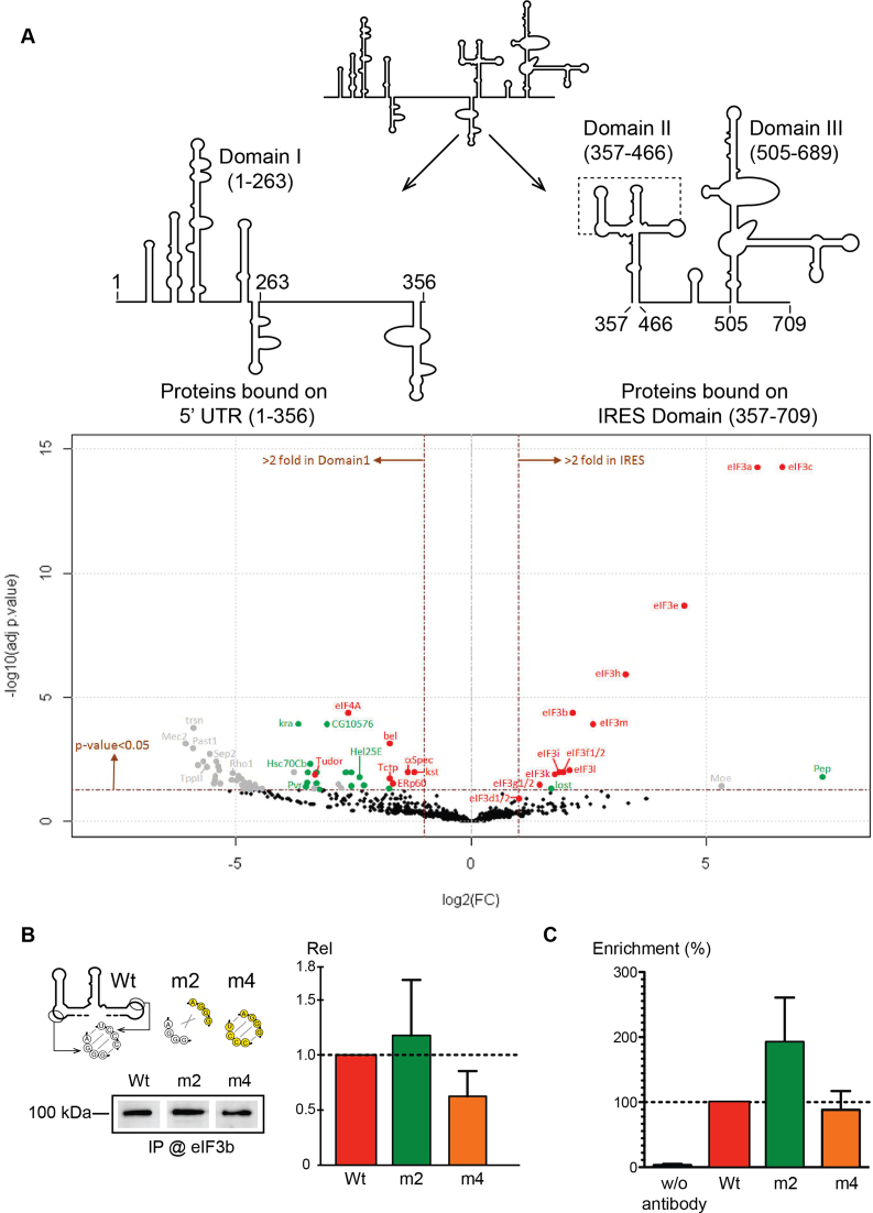
Cricket paralysis virus (CrPV) is a dicistrovirus. Its positive-sense single-stranded RNA genome contains two internal ribosomal entry sites (IRESs). The 5' untranslated region (5'UTR) IRES5'UTR mediates translation of non-structural proteins encoded by ORF1 whereas the well-known intergenic region (IGR) IRESIGR is required for translation of structural proteins from open reading frame 2 in the late phase of infection. Concerted action of both IRES is essential for host translation shut-off and viral translation. IRESIGR has been extensively studied, in contrast the IRES5'UTR remains largely unexplored. Here, we define the minimal IRES element required for efficient translation initiation in drosophila S2 cell-free extracts. We show that IRES5'UTR promotes direct recruitment of the ribosome on the cognate viral AUG start codon without any scanning step, using a Hepatitis-C virus-related translation initiation mechanism. Mass spectrometry analysis revealed that IRES5'UTR recruits eukaryotic initiation factor 3, confirming that it belongs to type III class of IRES elements. Using Selective 2'-hydroxyl acylation analyzed by primer extension and DMS probing, we established a secondary structure model of 5'UTR and of the minimal IRES5'UTR. The IRES5'UTR contains a pseudoknot structure that is essential for proper folding and ribosome recruitment. Overall, our results pave the way for studies addressing the synergy and interplay between the two IRES from CrPV.


Figures

Figure 1.
Figure 1.
Mapping of the minimal IRES sequence in the 5′UTR of CrPV by 5′ and 3′ truncation. On the left panels, a cartoon representation of Renilla luciferase reporter transcripts used in S2 cell-free translation extracts is shown. On the right panels, translation efficiencies are represented as raw bioluminescence activity (Relative Light Units or RLU) for each transcript. Standard deviations or translational activity for each transcript are shown and calculated from three independent experiments. **P < 0.005 based on Student‘s t-test. RNA integrity was controlled by 4% denaturing polyacrylamide gel electrophoresis (PAGE) and Ethidium Bromide staining. (A) IRES mapping within the entire CrPV 5′UTR and (B) precise mapping of 5′ and 3′ ends of the IRES5′UTR from CrPV.
Figure 2.
Figure 2.
AUG start codon recognition during CrPV IRES5′UTR-driven translation initiation Renilla luciferase reporter transcripts used in S2 cell-free translation are represented as raw bioluminescence activity (RLU) for each transcript. Standard deviations or translational activity for each transcript are shown and calculated from three independent experiments. **P < 0.005 based on Student’s t-test; ns, nonsignificant. RNA integrity was controlled by 4% denaturing PAGE and Ethidium Bromide staining. Viral coding sequences are shown in gray and fused to Renilla luciferase coding sequence. (A) IRES5′UTR drives translation initiation on viral cognate AUG start codon but does not promote scanning further downstream to find the Renilla AUG when the viral AUG is mutated to ACG (B) In these transcripts, the cognate viral AUG start codon is mutated to ACG. In addition in-frames AUGs were inserted at codon position 5 and 8 with the same context than the wt viral AUG. The 5′ proximal AUG codons are shown in bold. (C) In these transcripts the cognate viral AUG is shifted by 1, 2 or 3 nts.
Figure 3.
Figure 3.
The predicted secondary structure of the IRES5′UTR of CrPV reveals three highly structured domains named I, II and III separated by flexible linkers. Selective 2′-hydroxyl acylation analyzed by primer extension (SHAPE) data are overlaid on the structure prediction for the full-length 5′UTR RNA. Reactivities are shown as averages from three independent experiments (except for region 711–754: average from two experiments). Position of the minimal IRES is indicated (375–709). DMS and CMCT reactivity data for the minimal IRES domain (357–710). Reactivities are represented as averages from three independent experiments in the box of the right part of the figure. Nucleotides from J3/3a and L5 involved in long-distance interaction to form the pseudoknot are boxed. Reactivity values with standard deviations are listed in Supplementary Tables S1–4.
Figure 4.
Figure 4.
A pseudoknot is required for correct folding and efficient IRES5′UTR -driven translation. (A) Mutants in the J3/3a and/or L5 regions used in this study, mutated nucleotides are shown in yellow. (B) Absolute SHAPE reactivities for 357–754 RNA transcripts with either wt or m1–m4 mutant sequence. Reactivities are shown as averages from two independent experiments (reactivity values with standard deviation are given in Supplementary Table S2). The Pearson correlation coefficients (RPearson) with the 357–754 wt dataset are shown on the left of each histogram. J3/3a and L5 are boxed in green. (C) Translation activity of IRES 357–754 wt and mutants m1–m4 when placed upstream of Renilla luciferase coding region. RNA integrity control by denaturing 4% sodium dodecyl sulphate-PAGE is shown under the histogram. (D) Pre-initiation complex assembly and analysis on 7–47% sucrose gradient with P32 radio-labeled wt and mutant m1–m4 IRES. (E) In vivo translation assay using monocistronic reporters transfected in S2 cells. The mutants m1–m4 are compared with wt IRES5′UTR activity. *P < 0.05 and **P < 0.005, ***P < 0.0005 based on Student’s t-test.
Figure 5.
Figure 5.
Eukaryotic initiation factor (eIF)3 is recruited specifically by the IRES5′UTR in a pseudoknot-independent manner. (A) Mass spectrometry analysis of edeine-blocked translation initiation complexes on the 5′ proximal fragment (1–356) and the minimal IRES (357–709). Graphical representation of proteomics data: protein log2 spectral count fold changes (on the x-axis) and the corresponding adjusted log10P-values (on the y-axis) are plotted in a pairwise volcano plot. The significance thresholds are represented by a horizontal dashed line (P-value = 0.05, negative-binomial test with Benjamini–Hochberg adjustment) and two vertical dashed lines (−2.0-fold on the left and +2.0-fold on the right). Data points in the upper left and upper right quadrants indicate significant negative and positive changes in protein abundance. Protein names are labeled next to the off-centered spots and they are depicted according to the following color code: proteins represented by a red spot are identified by more than 30 MS/MS spectra, by a green spot when identified by 11–30 spectra and by a gray spot when identified by <10 spectra. Data points are plotted on the basis of average spectral counts from triplicate analysis. For an exhaustive list of hits, see Supplementary Table S5. (B) Western blots analysis with drosophila eIF3b antibody of edeine-blocked translation initiation complexes programmed with Wt and m2 and m4 mutants. The complexes were assembled in vitro in S2-cell extracts. The histogram represents the quantification of three independent experiments. (C) In vivo RNA immunoprecipitation of IRES5′UTR-Renilla reporter mRNA in S2 cells. Wt, m2 and m4 mRNA were immunoprecipitated using drosophila eIF3b antibody and quantified by RT-qPCR. The histogram represents the quantification of three independent experiments. The enrichment fold was calculated by the ratio between immunoprecipitated and the input and was set to 100% for the Wt.

References

    1. Walsh D., Mohr I.. Viral subversion of the host protein synthesis machinery. Nat. Rev. Microbiol. 2011; 9:860–875. - PMC - PubMed
    1. Joachims M., Van Breugel P.C., Lloyd R.E.. Cleavage of poly(A)-binding protein by enterovirus proteases concurrent with inhibition of translation in vitro. J. Virol. 1999; 73:718–727. - PMC - PubMed
    1. Etchison D., Milburn S.C., Edery I., Sonenberg N., Hershey J.W.. Inhibition of HeLa cell protein synthesis following poliovirus infection correlates with the proteolysis of a 220, 000-dalton polypeptide associated with eucaryotic initiation factor 3 and a cap binding protein complex. J. Biol. Chem. 1982; 257:14806–14810. - PubMed
    1. Kuyumcu-Martinez N.M., Joachims M., Lloyd R.E.. Efficient cleavage of ribosome-associated poly(A)-binding protein by enterovirus 3C protease. J. Virol. 2002; 76:2062–2074. - PMC - PubMed
    1. Jackson R.J., Hellen C.U.T., Pestova T. V. The mechanism of eukaryotic translation initiation and principles of its regulation. Nat. Rev. Mol. Cell Biol. 2010; 11:113–127. - PMC - PubMed

MeSH terms