. 2018 Jan:57:21-29.
doi: 10.1016/j.exphem.2017.09.003. Epub 2017 Sep 11.

Enhancement of mouse hematopoietic stem/progenitor cell function via transient gene delivery using integration-deficient lentiviral vectors

Affiliations

Enhancement of mouse hematopoietic stem/progenitor cell function via transient gene delivery using integration-deficient lentiviral vectors

Maria E Alonso-Ferrero et al. Exp Hematol. 2018 Jan.

Abstract

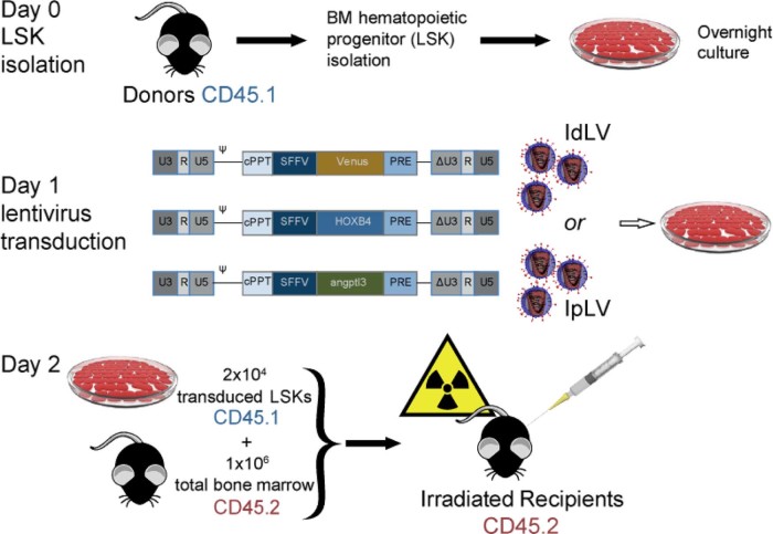
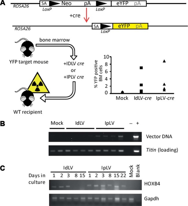
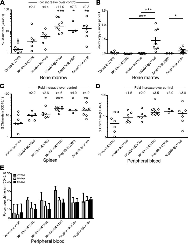
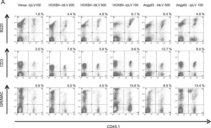
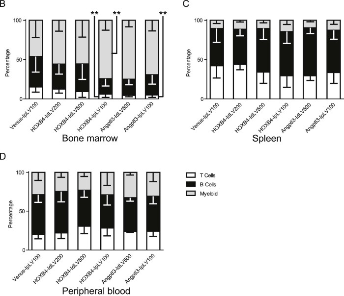
Integration-deficient lentiviruses (IdLVs) deliver genes effectively to tissues but are lost rapidly from dividing cells. This property can be harnessed to express transgenes transiently to manipulate cell biology. Here, we demonstrate the utility of short-term gene expression to improve functional potency of hematopoietic stem and progenitor cells (HSPCs) during transplantation by delivering HOXB4 and Angptl3 using IdLVs to enhance the engraftment of HSPCs. Constitutive overexpression of either of these genes is likely to be undesirable, but the transient nature of IdLVs reduces this risk and those associated with unsolicited gene expression in daughter cells. Transient expression led to increased multilineage hematopoietic engraftment in in vivo competitive repopulation assays without the side effects reported in constitutive overexpression models. Adult stem cell fate has not been programmed previously using IdLVs, but we demonstrate that these transient gene expression tools can produce clinically relevant alterations or be applied to investigate basic biology.


Figures

Figure 1
Figure 1
The in vivo competitive repopulation assay. BM cells were obtained by flushing femurs and tibias of C57BL/6J CD45.1 mice before staining with the lineage antigens CD3, CD45R (B220), CD11b, Gr-1 (Ly-6G/C), 7–4, and Ter-119. Lin cells were isolated using the mouse Lineage Cell Depletion Kit (Miltenyi Biotec) following the manufacturer's recommendations. Lin isolated cells were stained with streptavidin-FITC, PE Ly-6A/E (Sca-1), and APC CD117 (c-kit) (all obtained from BD Biosciences), and the LSK population was isolated in a MoFlo XDP sorter (Beckman Coulter). Cells were resuspended at 5 × 105 cells/mL in StemSpan SFEM (StemCell Technologies) supplemented with 0.5% penicillin–streptomycin, mSCF (300 ng/mL), hTPO (100 ng/mL, R&D Systems), and FLT3-L (100 ng/mL; Miltenyi Biotec) and cultured overnight before and after overnight culture and transduced for 24 hours with IdLVs or IpLVs at different MOIs. IdLV or IpLV versions of lentiviral vectors expressing a HOXB4 or Angptl3 or IpLV fluorophore (Venus, control) were used in this study. Self-inactivating vectors were produced with a ΔU3-deleted 3′ long-terminal repeat, with the transgene under the control of the internal spleen focus-forming virus long-terminal repeat promoter element. Transduced LSK cells were harvested the next day, and 2 × 104 of them were injected into the tail veins of lethally irradiated C57BL/6J CD45.2 recipient mice (split dose of 6 Gy and 4 Gy over 2 consecutive days, gamma irradiated using a caesium-137 source, dose rate 3 Gy/minute). One million freshly harvested CD45.2 total BM cells were injected alongside the CD45.1 donor LSKs as competitors. cPPT = Central polypurine tract; IRES = internal ribosomal entry site; PRE = attenuated post-transcriptional regulatory element; U5, R, and (Δ)U3 = (self-inactivating) long-terminal repeats; Ψ = packaging signal.
Figure 2
Figure 2
IdLV transduction of BM cells provides transient gene expression in vitro and in vivo. LSK cells were isolated from BM harvested from ROSA26-eYFP mice that contain an eYFP gene at the ROSA26 locus that is expressed upon exposure to cre-recombinase, which removes a disruptive neomycin (Neo) cassette . Cells were transduced with IdLVs or IpLVs encoding cre recombinase at an MOI of 100 before injecting into lethally irradiated wild-type (YFP-negative) recipients. (A) Analysis of recipient BM 6 months later revealed YFP-positive cells engrafted after cre recombinase expression from IdLVs and IpLVs. (B) Vector-specific PCR confirmed the long-term persistence for vector in IpLV samples, but not IdLV. (C) Lin cells were transduced with IdLVs or IpLVs expressing HOXB4 and grown in liquid culture. RT-PCR showed that HOXB4 expression from IdLVs can be observed for up to 3 days when cells are in culture, whereas IpLVs expressed the gene until the end of the experiment (22 days). Primers specific for gapdh were used as a positive control for the RT-PCR. blank = Non-template PCR control; mock = mock-transduced cells.
Figure 3
Figure 3
Transient delivery of HOXB4 and Angptl3 increases long-term donor cell chimerism in BM, spleen, and peripheral blood cells. IpLVs or IdLVs expressing HOXB4 or Angptl3 at different MOIs (100–500) or a control IpLV expressing the fluorophore Venus were used to transduce CD45.1 donor cells, which were transplanted into CD45.2 mice in an in vivo competitive repopulation assay. (A) BM cells from CD45.2 recipients were analyzed by flow cytometry to determine chimerism of donor CD45.1 cells at 90 days after injection. There was a statistical difference between samples over the whole group for BM (p < 0.0001, percentage data normalized by inverse-sine transformation followed by one-way analysis of variance [ANOVA]). *p < 0.5, **p < 0.01, ***p < 0.001 (Šidák's post hoc multiple comparison test vs. Venus IpLV 100). Bar shows the median value with fold increase in median values of samples compared with the control (top). (B) Real-time PCR vector copy number analysis in BM cells after 90 days. Error bars indicate mean value ±95% confidence intervals. Overall p < 0.0001, one-way ANOVA of copy numbers normalized by log transformation; *p < 0.5, ***p < 0.001, Šidák's post hoc multiple-comparisons test for comparisons between HOXB4 vectors and, separately, between Angptl3-treated mice. (C) Spleen and (D) peripheral blood from CD45.2 recipients were analyzed by flow cytometry to determine chimerism of transduced donor cells. There was a statistical difference between samples over the whole group for spleen cells collected at 90 days, p < 0.0001. Differences in chimerism in peripheral blood between groups did not reach statistical significance, except for HOXB4-IpLV100 versus Venus-IpLV100 (percentage data normalized by inverse-sine transformation followed by one-way ANOVA). *p < 0.5, **p < 0.01, ***p < 0.001, Šidák's post hoc multiple-comparisons test versus Venus-IpLV 100. (E) CD45.1 chimerism in the peripheral blood was measured at three time points over the 90-day period. Data shown are the median value; error bars indicate interquartile range.
Figure 4
Figure 4
Expression of HOXB4 or Angptl3 from IdLV does not skew lineage differentiation in transduced cells. (A) Representative flow cytometry plots showing levels of CD45.1 donor-derived T (CD3), B (B220), and myeloid (GR/MAC) cells (upper right quadrant) measured in recipient mouse PBMCs at 90 days after transplantation. The proportion that each lineage contributes to the average total CD45.1 cell population in each group was calculated by flow cytometry analysis in cells collected from (B) BM, (C) spleen, and (D) peripheral blood. One-way ANOVA on inverse sine-transformed percentage data for each lineage within each tissue reached significance (p < 0.05) for T-cell and myeloid lineages within BM cells. HOXB4-IpLV100 samples were significantly different from Venus IpLV-100 samples (p < 0.01) in the myeloid and T-cell lineages, and Angptl3-IpLV100 had a significantly lower proportion of T cells than the control (p < 0.01, Dunnett's post hoc multiple-comparisons test using a 99% confidence interval). n ≥ 3 in each group. Error bars indicate standard deviation.
Figure 4
Figure 4
Expression of HOXB4 or Angptl3 from IdLV does not skew lineage differentiation in transduced cells. (A) Representative flow cytometry plots showing levels of CD45.1 donor-derived T (CD3), B (B220), and myeloid (GR/MAC) cells (upper right quadrant) measured in recipient mouse PBMCs at 90 days after transplantation. The proportion that each lineage contributes to the average total CD45.1 cell population in each group was calculated by flow cytometry analysis in cells collected from (B) BM, (C) spleen, and (D) peripheral blood. One-way ANOVA on inverse sine-transformed percentage data for each lineage within each tissue reached significance (p < 0.05) for T-cell and myeloid lineages within BM cells. HOXB4-IpLV100 samples were significantly different from Venus IpLV-100 samples (p < 0.01) in the myeloid and T-cell lineages, and Angptl3-IpLV100 had a significantly lower proportion of T cells than the control (p < 0.01, Dunnett's post hoc multiple-comparisons test using a 99% confidence interval). n ≥ 3 in each group. Error bars indicate standard deviation.

Similar articles

Cited by

References

    1. Milanovich S., Peterson J., Allred J. Sall4 overexpression blocks murine hematopoiesis in a dose-dependent manner. Exp Hematol. 2015;43:53–64. e1-e8. - PMC - PubMed
    1. Sakuma T., Barry M.A., Ikeda Y. Lentiviral vectors: basic to translational. Biochem J. 2012;443:603–618. - PubMed
    1. Aiuti A., Biasco L., Scaramuzza S. Lentiviral hematopoietic stem cell gene therapy in patients with Wiskott-Aldrich syndrome. Science. 2013;341:1233151. - PMC - PubMed
    1. Leavitt A.D., Robles G., Alesandro N., Varmus H.E. Human immunodeficiency virus type 1 integrase mutants retain in vitro integrase activity yet fail to integrate viral DNA efficiently during infection. J Virol. 1996;70:721–728. - PMC - PubMed
    1. Yáñez-Muñoz R.J., Balaggan K.S., MacNeil A. Effective gene therapy with nonintegrating lentiviral vectors. Nat Med. 2006;12:348–353. - PubMed

MeSH terms