

Mitotic transcription and waves of gene reactivation during mitotic exit
- PMID: 28912132
- PMCID: PMC5727891
- DOI: 10.1126/science.aal4671
Mitotic transcription and waves of gene reactivation during mitotic exit
Abstract
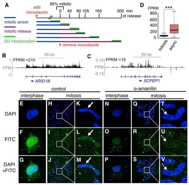
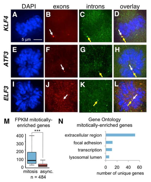
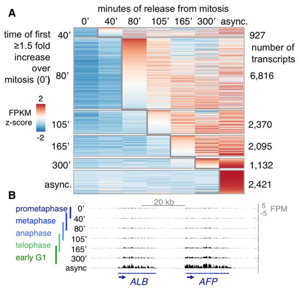
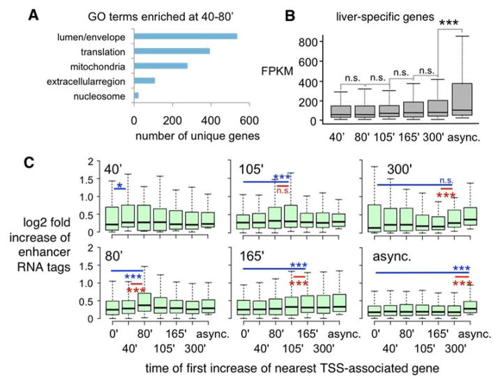
Although the genome is generally thought to be transcriptionally silent during mitosis, technical limitations have prevented sensitive mapping of transcription during mitosis and mitotic exit. Thus, the means by which the interphase expression pattern is transduced to daughter cells have been unclear. We used 5-ethynyluridine to pulse-label transcripts during mitosis and mitotic exit and found that many genes exhibit transcription during mitosis, as confirmed with fluorescein isothiocyanate-uridine 5'-triphosphate labeling, RNA fluorescence in situ hybridization, and quantitative reverse transcription polymerase chain reaction. The first round of transcription immediately after mitosis primarily activates genes involved in the growth and rebuilding of daughter cells, rather than cell type-specific functions. We propose that the cell's transcription pattern is largely retained at a low level through mitosis, whereas the amplitude of transcription observed in interphase is reestablished during mitotic exit.
Copyright © 2017 The Authors, some rights reserved; exclusive licensee American Association for the Advancement of Science. No claim to original U.S. Government Works.
Figures
Comment in
-
Transcription: No proper rest in mitosis.Nat Rev Mol Cell Biol. 2017 Nov;18(11):652-653. doi: 10.1038/nrm.2017.102. Epub 2017 Oct 4. Nat Rev Mol Cell Biol. 2017. PMID: 28974773 No abstract available.
-
Mitotic Chromosomes: Not So Silent After All.Dev Cell. 2017 Oct 23;43(2):119-121. doi: 10.1016/j.devcel.2017.10.002. Dev Cell. 2017. PMID: 29065303
References
-
- Antonin W, Neumann H. Chromosome condensation and decondensation during mitosis. Curr Opin Cell Biol. 2016;40:15–22. - PubMed
-
- Gottesfeld JM, Forbes DJ. Mitotic repression of the transcriptional machinery. Trends Biochem Sci. 1997;22:197–202. - PubMed
-
- Martinez-Balbas MA, Dey A, Rabindran SK, Ozato K, Wu C. Displacement of sequence-specific transcription factors from mitotic chromatin. Cell. 1995;83:29–38. - PubMed
Publication types
MeSH terms
Substances
Grants and funding
LinkOut - more resources
Full Text Sources
Other Literature Sources
Molecular Biology Databases
