

Review
doi: 10.1161/JAHA.117.006339.
Valve Interstitial Cells: The Key to Understanding the Pathophysiology of Heart Valve Calcification
Affiliations
- PMID: 28912209
- PMCID: PMC5634284
- DOI: 10.1161/JAHA.117.006339
Item in Clipboard
Review
Valve Interstitial Cells: The Key to Understanding the Pathophysiology of Heart Valve Calcification
J Am Heart Assoc.
.
No abstract available
Keywords: aortic stenosis; ectopic bone formation; myofibroblasts; valve endothelial cells; valve interstitial cells.
Figures
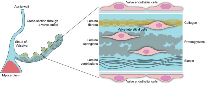
Simplified structure of the human aortic valve leaflet. On the left is a schematic cross section through the noncoronary leaflet of the aortic valve. The blowup on the right shows the trilayered organization of the extracellular matrix and the localization of the aortic valve endothelial cells (shortened throughout to VEC s) and interstitial cells (VIC s).
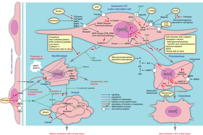
A current understanding of the pathological differentiation of valvular interstitial cells in aortic valve calcification. The cell types are given in blue. Quiescent valvular interstitial cels (VIC s) as an effect of exogenous stimuli (given in yellow fields) differentiate into myofibroblasts (left) or preosteoblasts (right). Differentiation process is shown as gray arrows. The myofibroblasts can further assemble themselves into nodules, which undergo apoptosis and provide substrate for diffuse calcification (bottom left). The process is negatively regulated by valve endothelial cells (far left). The preosteoblasts can further differentiate into osteoblasts, which in turn synthesize ordinary bone (bottom right). The processes are orchestrated by a complex network of factors. The ligands stimulate surface receptors (white circles), which further relay to the signaling networks (black arrows). The signals can be inhibitory (stump arrows) or stimulatory (arrows with a “+”). The signal can constitute stimulation of expression of certain factors, a process shown as blue arrows. Generally the procalcific stimuli are shown with red arrows, and anticalcific are given in green. Due to the scheme complexity, several factors appear in multiple places on the scheme. ALP indicates alkaline phosphatase; aSMA, α‐smooth muscle actin; BMP, bone morphogenetic protein; BMPR, bone morphogenetic protein receptor; DAPT, inhibitor of γ‐secretase; ENOS, endothelial nitric oxide synthase; ENPP, ectonucleotide pyrophosphatase/phosphodiesterase 1; ERK, extracellular signal‐regulated kinase; FGF, fibroblast growth factor; IL, interleukin; LPS, lipopolysaccharide; MAPK, mitogen‐associated protein kinase; MMP, matrix metalloproteinase; NFkB Nuclear factor κB; NICD, Notch intracellular domain; NO, nitric oxide; NotchR, Notch receptor; oxLDL, oxidized low‐density lipoproteids; PGN, peptidoglycan; Runx2, runt‐related transcriptional factor 2; SMAD, small mothers against decapentaplegic; TGF, transforming growth factor; TLR, toll‐like receptor; TNF, tumor necrosis factor.
References
-
- Iung B, Baron G, Butchart EG, Delahaye F, Gohlke‐Bärwolf C, Levang OW, Tornos P, Vanoverschelde J‐L, Vermeer F, Boersma E. A prospective survey of patients with valvular heart disease in Europe: the Euro Heart Survey on Valvular Heart Disease. Eur Heart J. 2003;24:1231–1243. - PubMed
-
- O'Brien KD. Epidemiology and genetics of calcific aortic valve disease. J Investig Med. 2007;55:284–291. - PubMed
-
- Goldbarg SH, Elmariah S, Miller MA, Fuster V. Insights into degenerative aortic valve disease. J Am Coll Cardiol. 2007;50:1205–1213. - PubMed
Publication types
MeSH terms
Substances
Supplementary concepts
LinkOut - more resources
Full Text Sources
Other Literature Sources
