

Using a Novel Microfabricated Model of the Alveolar-Capillary Barrier to Investigate the Effect of Matrix Structure on Atelectrauma
- PMID: 28912466
- PMCID: PMC5599538
- DOI: 10.1038/s41598-017-12044-9
Using a Novel Microfabricated Model of the Alveolar-Capillary Barrier to Investigate the Effect of Matrix Structure on Atelectrauma
Abstract
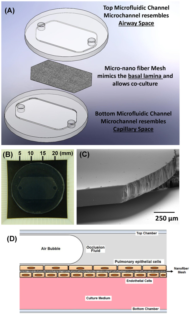
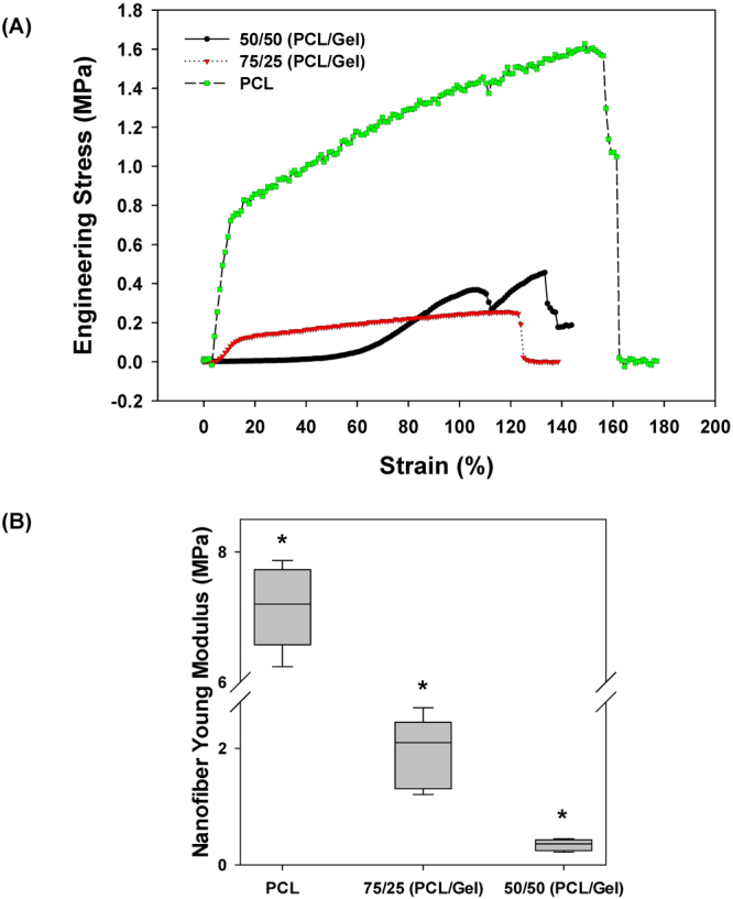
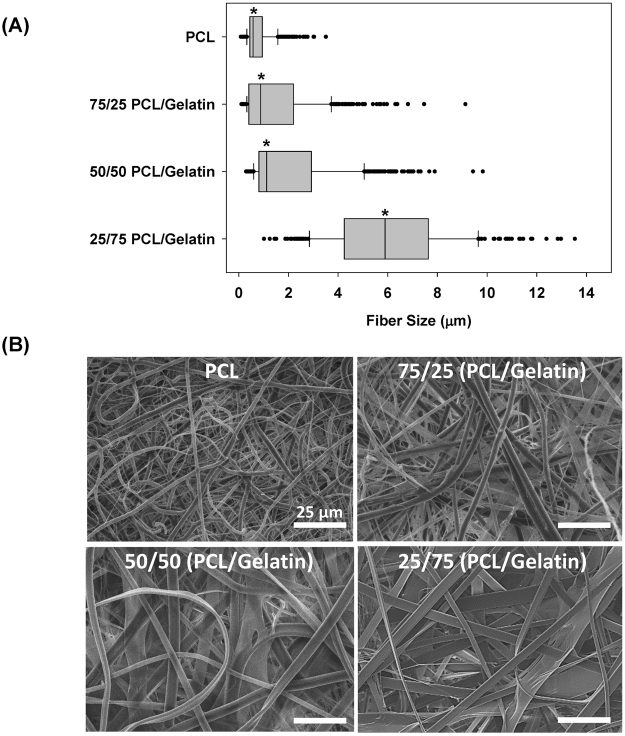
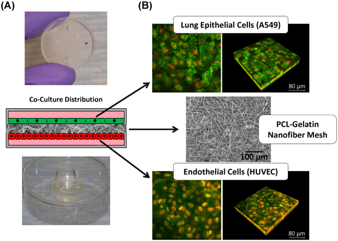
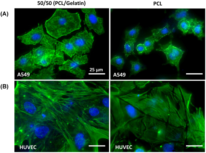
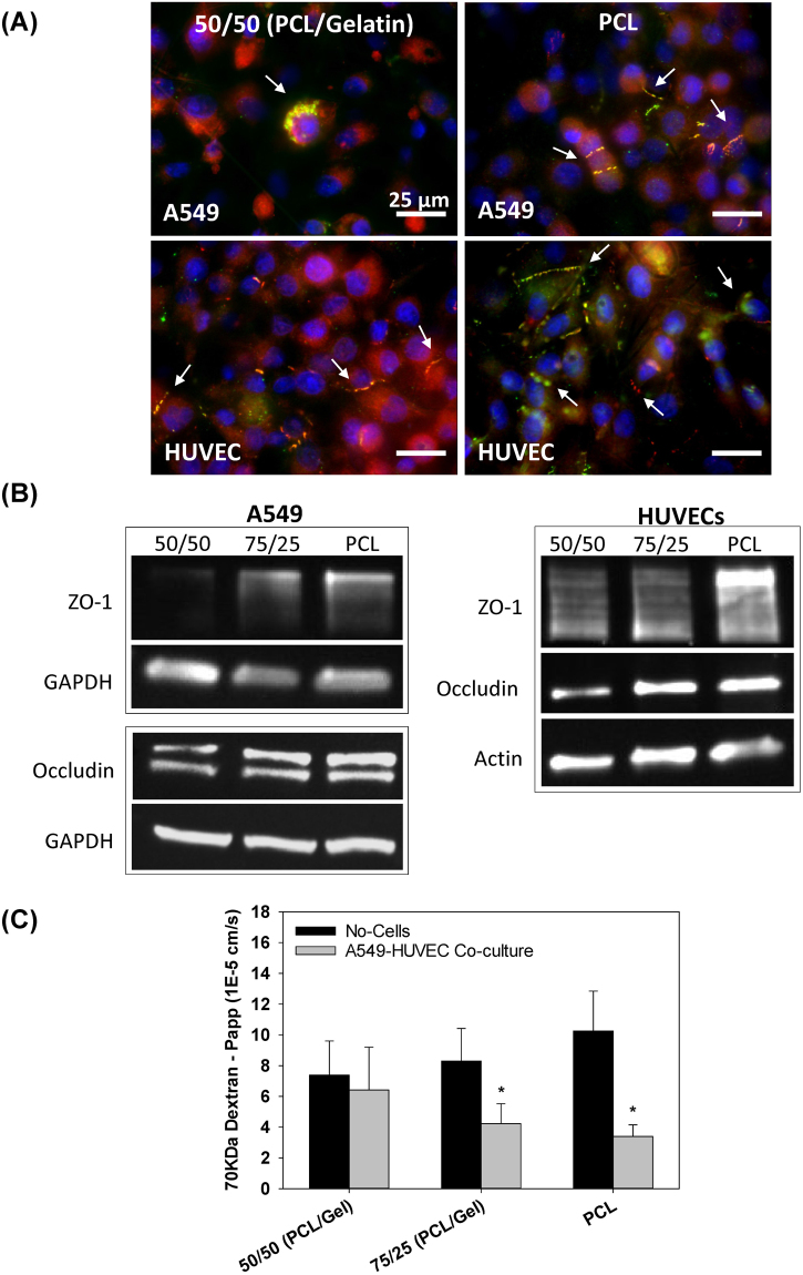
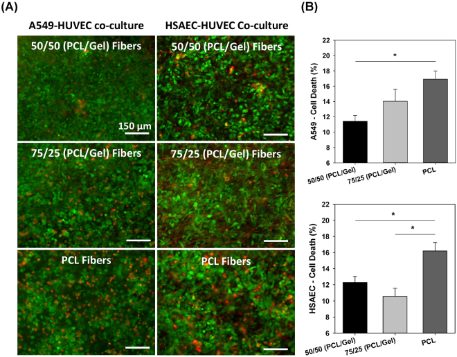
The alveolar-capillary barrier is composed of epithelial and endothelial cells interacting across a fibrous extracelluar matrix (ECM). Although remodeling of the ECM occurs during several lung disorders, it is not known how fiber structure and mechanics influences cell injury during cyclic airway reopening as occurs during mechanical ventilation (atelectrauma). We have developed a novel in vitro platform that mimics the micro/nano-scale architecture of the alveolar microenvironment and have used this system to investigate how ECM microstructural properties influence epithelial cell injury during airway reopening. In addition to epithelial-endothelial interactions, our platform accounts for the fibrous topography of the basal membrane and allows for easy modulation of fiber size/diameter, density and stiffness. Results indicate that fiber stiffness and topography significantly influence epithelial/endothelial barrier function where increased fiber stiffness/density resulted in altered cytoskeletal structure, increased tight junction (TJ) formation and reduced barrier permeability. However, cells on rigid/dense fibers were also more susceptible to injury during airway reopening. These results indicate that changes in the mechanics and architecture of the lung microenvironment can significantly alter cell function and injury and demonstrate the importance of implementing in vitro models that more closely resemble the natural conditions of the lung microenvironment.
Conflict of interest statement
The authors declare that they have no competing interests.
Figures
Similar articles
-
Human alveolar epithelial cells expressing tight junctions to model the air-blood barrier.ALTEX. 2016;33(3):251-60. doi: 10.14573/altex.1511131. Epub 2016 Mar 17. ALTEX. 2016. PMID: 26985677
-
Differential regulation of human lung epithelial and endothelial barrier function by thrombin.Am J Respir Cell Mol Biol. 2004 Nov;31(5):517-27. doi: 10.1165/rcmb.2003-0432OC. Epub 2004 Jul 29. Am J Respir Cell Mol Biol. 2004. PMID: 15284075
-
Influence of cytoskeletal structure and mechanics on epithelial cell injury during cyclic airway reopening.Am J Physiol Lung Cell Mol Physiol. 2009 Nov;297(5):L881-91. doi: 10.1152/ajplung.90562.2008. Epub 2009 Aug 21. Am J Physiol Lung Cell Mol Physiol. 2009. PMID: 19700641
-
The blood-gas barrier in COVID-19: an overview of the effects of SARS-CoV-2 infection on the alveolar epithelial and endothelial cells of the lung.Tissue Barriers. 2021 Oct 2;9(4):1937013. doi: 10.1080/21688370.2021.1937013. Epub 2021 Jul 7. Tissue Barriers. 2021. PMID: 34232823 Free PMC article. Review.
-
Alveolar epithelial barrier and acute lung injury.New Horiz. 1993 Nov;1(4):613-22. New Horiz. 1993. PMID: 8087581 Review.
Cited by
-
A Robust Protocol for Decellularized Human Lung Bioink Generation Amenable to 2D and 3D Lung Cell Culture.Cells. 2021 Jun 18;10(6):1538. doi: 10.3390/cells10061538. Cells. 2021. PMID: 34207111 Free PMC article.
-
Simple-Flow: A 3D-Printed Multiwell Flow Plate to Coculture Primary Human Lung Cells at the Air-Liquid Interface.ACS Biomater Sci Eng. 2025 Jan 13;11(1):451-462. doi: 10.1021/acsbiomaterials.4c01322. Epub 2024 Dec 24. ACS Biomater Sci Eng. 2025. PMID: 39719361
-
A miniaturized multicellular platform to mimic the 3D structure of the alveolar-capillary barrier.Front Bioeng Biotechnol. 2024 Apr 5;12:1346660. doi: 10.3389/fbioe.2024.1346660. eCollection 2024. Front Bioeng Biotechnol. 2024. PMID: 38646009 Free PMC article.
-
Protective Effects of Kirenol against Lipopolysaccharide-Induced Acute Lung Injury through the Modulation of the Proinflammatory NFκB Pathway and the AMPK2-/Nrf2-Mediated HO-1/AOE Pathway.Antioxidants (Basel). 2021 Jan 31;10(2):204. doi: 10.3390/antiox10020204. Antioxidants (Basel). 2021. PMID: 33572510 Free PMC article.
-
Nanoparticle delivery of microRNA-146a regulates mechanotransduction in lung macrophages and mitigates injury during mechanical ventilation.Nat Commun. 2021 Jan 12;12(1):289. doi: 10.1038/s41467-020-20449-w. Nat Commun. 2021. PMID: 33436554 Free PMC article.
References
-
- Vlahakis NE, Schroeder MA, Limper AH, Hubmayr RD. Stretch induces cytokine release by alveolar epithelial cells in vitro. Am J Physiol. 1999;277:L167–173. - PubMed
Publication types
MeSH terms
Substances
Grants and funding
LinkOut - more resources
Full Text Sources
Other Literature Sources
Medical
