. 2017 Sep 14;7(1):11541.
doi: 10.1038/s41598-017-11177-1.

Transcription regulation of CDKN1A (p21/CIP1/WAF1) by TRF2 is epigenetically controlled through the REST repressor complex

Affiliations

Transcription regulation of CDKN1A (p21/CIP1/WAF1) by TRF2 is epigenetically controlled through the REST repressor complex

Tabish Hussain et al. Sci Rep. .

Abstract

We observed extra-telomeric binding of the telomere repeat binding factor TRF2 within the promoter of the cyclin-dependent kinase CDKNIA (p21/CIP1/WAF1). This result in TRF2 induced transcription repression of p21. Interestingly, p21 repression was through engagement of the REST-coREST-LSD1-repressor complex and altered histone marks at the p21 promoter in a TRF2-dependent fashion. Furthermore, mutational analysis shows p21 repression requires interaction of TRF2 with a p21 promoter G-quadruplex. Physiologically, TRF2-mediated p21 repression attenuated drug-induced activation of cellular DNA damage response by evading G2/M arrest in cancer cells. Together these reveal for the first time role of TRF2 in REST- repressor complex mediated transcription repression.


Conflict of interest statement

The authors declare that they have no competing interests.

Figures

Figure 1
Figure 1
TRF2 transcriptionally regulates p21 expression through promoter occupancy. (A) Quantitative ChIP using TRF2 antibody gives enriched binding of TRF2 on p21 promoter in HT1080 and MDA-MB-231 cells, IgG was used as isotypic antibody control; normalized with 10% input (data represented as mean ± SEM, for three replicates). (B,C) TRF2 represses p21 promoter activity. In luciferase assay, si-RNA-mediated silencing or TRF2 over expression resulted in increased (B, *p value < 0.05, Student’s t-test; data represented as mean ± SEM of three replicates) or reduction (C, *p value < 0.05, Student’s t-test; data represented as mean ± SEM of three replicates) in p21 promoter activity in HT1080 and MDA-MB-231 cells, respectively; over expression of TRF2 devoid of DNA binding [deletion of basic (delB), myb (delM) and both basic/myb (delB-delM) domains] resulted in partial or complete rescue of p21 promoter activity in HT1080 and MDA-MB-231 cells (data represented as mean ± SEM, for three replicates) (C). (D,E) HT1080 cells over expressing TRF2 show reduced p21 protein expression, bar graph shows the densitometry analysis of three different immunoblot replicates (*p value < 0.05, Student’s t-test; data represented as mean ± SEM of three replicates). (E) (Full images are shown in Supplementary Figure S5); TRF2 silencing results in increase in p21 protein, bar graph shows the densitometry analysis of three different immunoblots (*p value < 0.05, Student’s t-test; data represented as mean ± SEM of three replicates) (F); and mRNA expression in HT1080 cells (*p value < 0.05, Student’s t-test; data represented as mean ± SEM of three replicates, GAPDH used as internal control for real-time PCR).
Figure 2
Figure 2
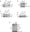
Repressive chromatin induced on p21 promoter in TRF2 dependent way. (AC) Loss of chromatin activation marks H3K4Me, H3K4Me2 in stable TRF2 expressed HT1080 cells (A); increase in LSD1 occupancy in TRF2 over-expressing HT1080 cells. (B) (Full images are shown in Supplementary Figures S6 and S7); Co-immunoprecipitation of TRF2 with LSD1 (immunoprecipitation with anti-TRF2 antibody followed by immunoblotting with anti-LSD1 or anti-TRF2 antibody) (C); (D) Reverse Co-immunoprecipitation of LSD1 with TRF2 (immunoprecipitation with anti-LSD1 antibody followed by immunoblotting with anti-TRF2 antibody); (E) and reduced occupancy of the repressor complex REST, Co-REST and LSD1 on silencing TRF2 on p21 promoter.
Figure 3
Figure 3
TRF2 binding at p21 promoter and p21 promoter activity is G4-motif dependent. (A) p21 promoter showing position of G4-motif. (B) Circular dichroism showing oligonucleotide constituting potential G4-motif adopts G4 structure in solution and on substitution of bases (in red font) required for G4 formation (p21-G4mut1, p21-G4mut2, p21-G4mut3) gives partial/complete disruption of the G4-motif under similar conditions; (C) interaction of recombinant TRF2 with G4-motif, mutated p21-G4mut3 or double stranded oligonucleotide of identical sequence was checked using ELISA. (D) Disruption of promoter G4-motif rescues TRF2-mediated repression of p21 promoter activity. Base substitutions that give partial/complete disruption of p21 G4-motif (p21-G4mut1, p21-G4mut2, p21-G4mut3) attenuate TRF2-dependent promoter repression (*p value < 0.05, Student’s t-test; data represented as mean ± SEM of three replicates). (E) TRF2 ChIP following treatment with G4-motif-specific ligand 360A gave reduced TRF2 occupancy at the endogenous p21 promoter relative to DMSO treatment as control (Full image is shown in Supplementary Figure S8).
Figure 4
Figure 4
Transcriptional regulation of p21 by TRF2 is active in p53-depleted conditions. (AD) p21 activation on TRF2 suppression is significant in p53 knock down cells. TRF2 silencing results in p21 activation in HT1080 cells with endogenous p53 expression (column 1 and 2). On siRNA-meditated suppression of p53, p21 activation is TRF2 dependent–on TRF2 suppression p21 expression increases (column 4), relative to endogenous TRF2 levels (column 3) at protein (A) and mRNA level (error bars represent ± SEM, GAPDH used as internal control for real-time PCR; data represented as mean ± SEM, for three replicates) (B). TRF2 regulate p21 in HCT116 p53−/− cells. TRF2 silencing results in p21 activation in HCT116 p53−/− cells at protein (C) and mRNA level (B,D) data represented as mean ± SEM, for three replicates) (Full images are shown in Supplementary Figure S9).
Figure 5
Figure 5
TRF2 represses p21 activation upon cellular DNA damage. (AC) Treatment with either Doxorubicin or Topotecan was done in HT1080 (A), *p value < 0.05, Student’s t-test; data represented as mean ± SEM of three replicates), MDA-MB-231 (B), data represented as mean ± SEM, for three replicates) and HCT116 p53−/− (C), data represented as mean ± SEM, for three replicates) cells following over-expression of TRF2: loss in p21 activation relative to control vector transformed cells was observed in each case (GAPDH used as internal control for real-time PCR). (D) Quantitative ChIP result shows partial loss of TRF2 occupancy at p21 promoter after doxorubicin treatment in HT1080 cell line.
Figure 6
Figure 6
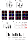
G4-binding ligand 360A rescues TRF2 mediated p21 suppression. (A,B) TRF2 mediated p21 repression is rescued by G4-binding ligands. A combination treatment with Doxorubicin and intracellular G4-ligand 360A rescues p21 activation relative to Doxorubicin or 360A treatment alone in HT1080 (A), *p value < 0.05, Student’s t-test; data represented as mean ± SEM of three replicates) and MDA-MB-231 cells (B), data represented as mean ± SEM, for three replicates) (GAPDH used as internal control for real-time PCR). (C) Relative loss in activation of p21 expression in cells over-expressing TRF2 relative to vector control cells was also observed by immunofluorescence in HT1080 cells on treatment with DMSO, and doxorubicin (first two panels). Treatment with 360A along with doxorubicin increased p21 activation (rightmost panel) (scale bar = 5 μm). (D,E) Increase in cells in G2/M phase in Doxorubicin + 360A combination treatment relative to treatment with Doxorubicin or 360A alone was observed (*p value < 0.05, Student’s t-test; data represented as mean ± SEM of three replicates). (F) Enhanced cytotoxic effect on cancer cells was seen with a combined treatment of Doxorubicin + 360A relative to treatment with Doxorubicin or 360A alone.

Similar articles

Cited by

References

    1. Greider CW. Telomeres, telomerase and senescence. BioEssays News Rev. Mol. Cell. Dev. Biol. 1990;12:363–369. doi: 10.1002/bies.950120803. - DOI - PubMed
    1. Blackburn EH. Telomeres: no end in sight. Cell. 1994;77:621–623. doi: 10.1016/0092-8674(94)90046-9. - DOI - PubMed
    1. Zakian VA. Telomeres: beginning to understand the end. Science. 1995;270:1601–1607. doi: 10.1126/science.270.5242.1601. - DOI - PubMed
    1. van Steensel B, Smogorzewska A, de Lange T. TRF2 protects human telomeres from end-to-end fusions. Cell. 1998;92:401–413. doi: 10.1016/S0092-8674(00)80932-0. - DOI - PubMed
    1. de Lange T. Shelterin: the protein complex that shapes and safeguards human telomeres. Genes Dev. 2005;19:2100–2110. doi: 10.1101/gad.1346005. - DOI - PubMed

Publication types

MeSH terms

Substances