

Cordycepin induces apoptosis of human acute monocytic leukemia cells via downregulation of the ERK/Akt signaling pathway
- PMID: 28912858
- PMCID: PMC5585717
- DOI: 10.3892/etm.2017.4855
Cordycepin induces apoptosis of human acute monocytic leukemia cells via downregulation of the ERK/Akt signaling pathway
Abstract
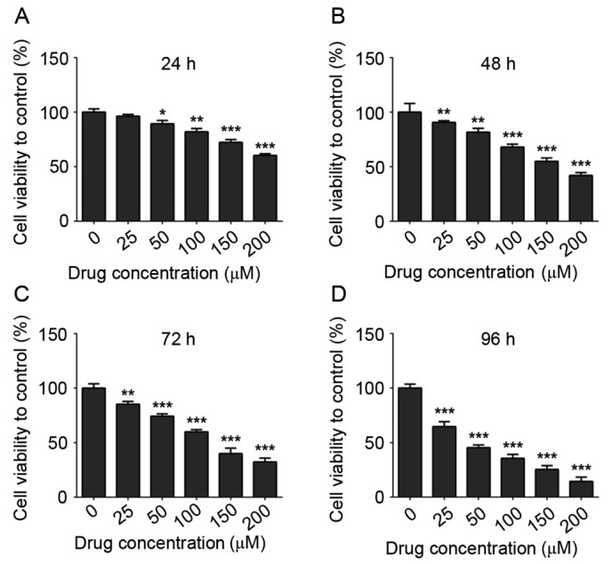
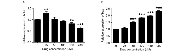
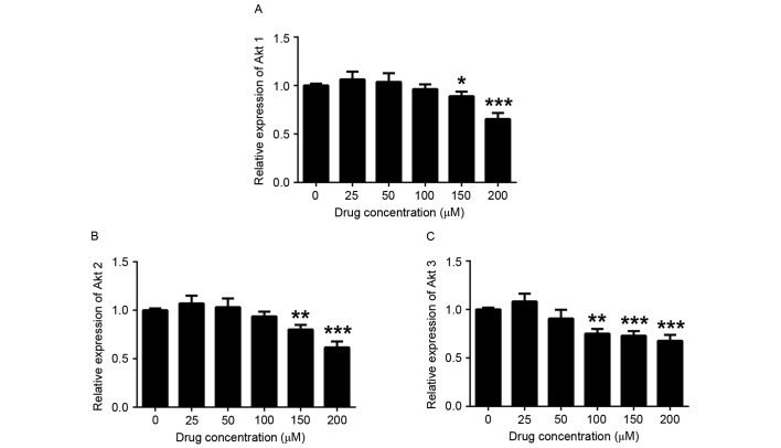
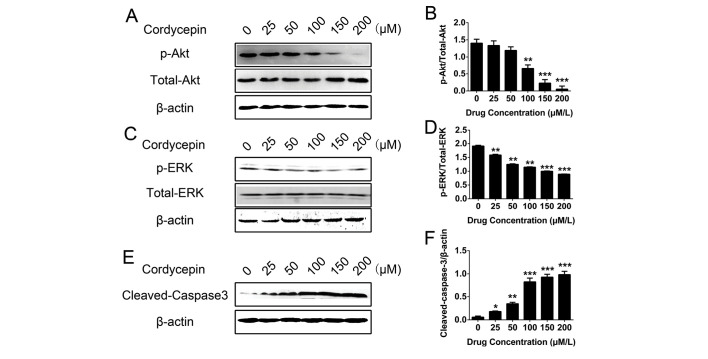
The aim of the present study was to examine the apoptotic effect of cordycepin (COR) on human THP-1 acute monocytic leukemia cells. THP-1 cells were exposed to different concentrations of COR for 24, 48, 72 or 96 h. The cell viability and apoptotic rate were analyzed. The gene expression of Akt1, Akt2, Akt3, B-cell lymphoma 2 (Bcl-2) and Bcl-2-associated X protein (Bax) were assessed by reverse-transcription quantitative PCR. Western blot analysis was used to detect the protein levels of phosphorylated (p)-Akt, p-extracellular signal-regulated kinase (ERK) and cleaved caspase-3. It was found that the viability of THP-1 cells was inhibited by COR in a dose- and time-dependent manner. After treatment with 200 µM COR for 24 h, the percentage of apoptotic cells was significantly increased. COR also downregulated the levels of Bcl-2, Akt1, Akt2 and Akt3, and elevated the expression of Bax. The protein levels of p-Akt and p-ERK were suppressed and cleaved caspase-3 was increased after treatment of COR. In conclusion, COR was found to induce apoptosis of THP-1 acute monocytic leukemia cells through downregulation of ERK/Akt signaling.
Keywords: Akt; ERK; THP-1; apoptosis; caspase-3; cordycepin.
Figures

Similar articles
-
Dihydroartemisinin-induced apoptosis in human acute monocytic leukemia cells.Oncol Lett. 2018 Mar;15(3):3178-3184. doi: 10.3892/ol.2017.7644. Epub 2017 Dec 19. Oncol Lett. 2018. PMID: 29435054 Free PMC article.
-
Cordycepin promotes apoptosis by modulating the ERK-JNK signaling pathway via DUSP5 in renal cancer cells.Am J Cancer Res. 2016 Aug 1;6(8):1758-71. eCollection 2016. Am J Cancer Res. 2016. PMID: 27648363 Free PMC article.
-
Curcumin induces apoptosis via simultaneously targeting AKT/mTOR and RAF/MEK/ERK survival signaling pathways in human leukemia THP-1 cells.Pharmazie. 2014 Mar;69(3):229-33. Pharmazie. 2014. PMID: 24716415
-
RKTG overexpression inhibits proliferation and induces apoptosis of human leukemia cells via suppression of the ERK and PI3K/AKT signaling pathways.Oncol Lett. 2017 Jul;14(1):965-970. doi: 10.3892/ol.2017.6182. Epub 2017 May 17. Oncol Lett. 2017. PMID: 28693259 Free PMC article.
-
Involvement of smad2 and Erk/Akt cascade in TGF-β1-induced apoptosis in human gingival epithelial cells.Cytokine. 2015 Sep;75(1):165-73. doi: 10.1016/j.cyto.2015.03.011. Epub 2015 Apr 14. Cytokine. 2015. PMID: 25882870
Cited by
-
Cordycepin (3'dA) Induces Cell Death of AC133+ Leukemia Cells via Re-Expression of WIF1 and Down-Modulation of MYC.Cancers (Basel). 2023 Aug 2;15(15):3931. doi: 10.3390/cancers15153931. Cancers (Basel). 2023. PMID: 37568748 Free PMC article.
-
[Effect of polydatin on the proliferation and apoptosis of THP-1 cells and the mechanism].Zhongguo Dang Dai Er Ke Za Zhi. 2022 Jul 15;24(7):821-825. doi: 10.7499/j.issn.1008-8830.2202078. Zhongguo Dang Dai Er Ke Za Zhi. 2022. PMID: 35894200 Free PMC article. Chinese.
-
A Systematic Review of the Biological Effects of Cordycepin.Molecules. 2021 Sep 28;26(19):5886. doi: 10.3390/molecules26195886. Molecules. 2021. PMID: 34641429 Free PMC article.
-
Sodium benzoate attenuates 2,8-dihydroxyadenine nephropathy by inhibiting monocyte/macrophage TNF-α expression.Sci Rep. 2023 Feb 27;13(1):3331. doi: 10.1038/s41598-023-30056-6. Sci Rep. 2023. PMID: 36849798 Free PMC article.
-
Cordycepin induces apoptosis in human pancreatic cancer cells via the mitochondrial-mediated intrinsic pathway and suppresses tumor growth in vivo.Onco Targets Ther. 2018 Aug 1;11:4479-4490. doi: 10.2147/OTT.S164670. eCollection 2018. Onco Targets Ther. 2018. PMID: 30122940 Free PMC article.
References
-
- Khaled S, Al Malki M, Marcucci G. Acute myeloid leukemia: Biologic, prognostic, and therapeutic insights. Oncology (Williston Park) 2016;30:318–329. - PubMed
-
- Pettersson L, Levéen P, Axler O, Dvorakova D, Juliusson G, Ehinger M. Improved minimal residual disease detection by targeted quantitative polymerase chain reaction in Nucleophosmin 1 type a mutated acute myeloid leukemia. Genes Chromosomes Cancer. 2016;55:750–766. doi: 10.1002/gcc.22375. - DOI - PubMed
-
- Brumatti G, Ma C, Lalaoui N, Nguyen NY, Navarro M, Tanzer MC, Richmond J, Ghisi M, Salmon JM, Silke N, et al. The caspase-8 inhibitor emricasan combines with the SMAC mimetic birinapant to induce necroptosis and treat acute myeloid leukemia. Sci Transl Med. 2016;8:339ra69. doi: 10.1126/scitranslmed.aad3099. - DOI - PubMed
LinkOut - more resources
Full Text Sources
Other Literature Sources
Research Materials
Miscellaneous