

The Effect of Er:YAG Laser Irradiation and Different Concentrations of Sodium Hypochlorite on Shear Bond Strength of Composite to Primary Teeth's Dentin
- PMID: 28912941
- PMCID: PMC5420362
- DOI: 10.15171/jlms.2017.06
The Effect of Er:YAG Laser Irradiation and Different Concentrations of Sodium Hypochlorite on Shear Bond Strength of Composite to Primary Teeth's Dentin
Abstract
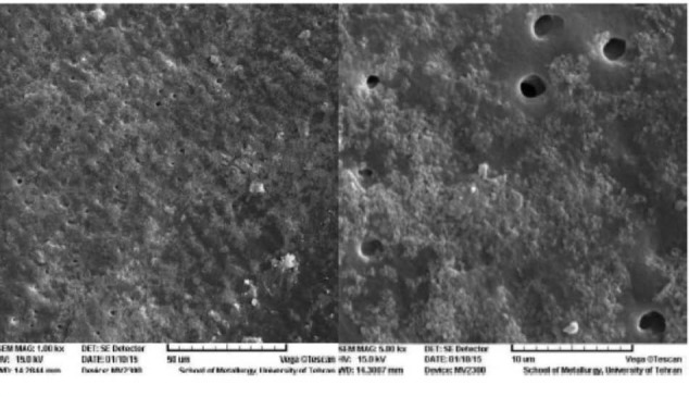
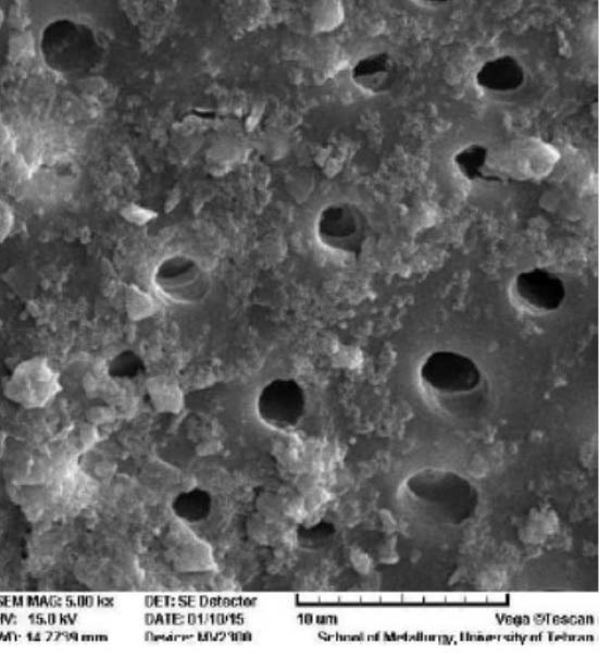
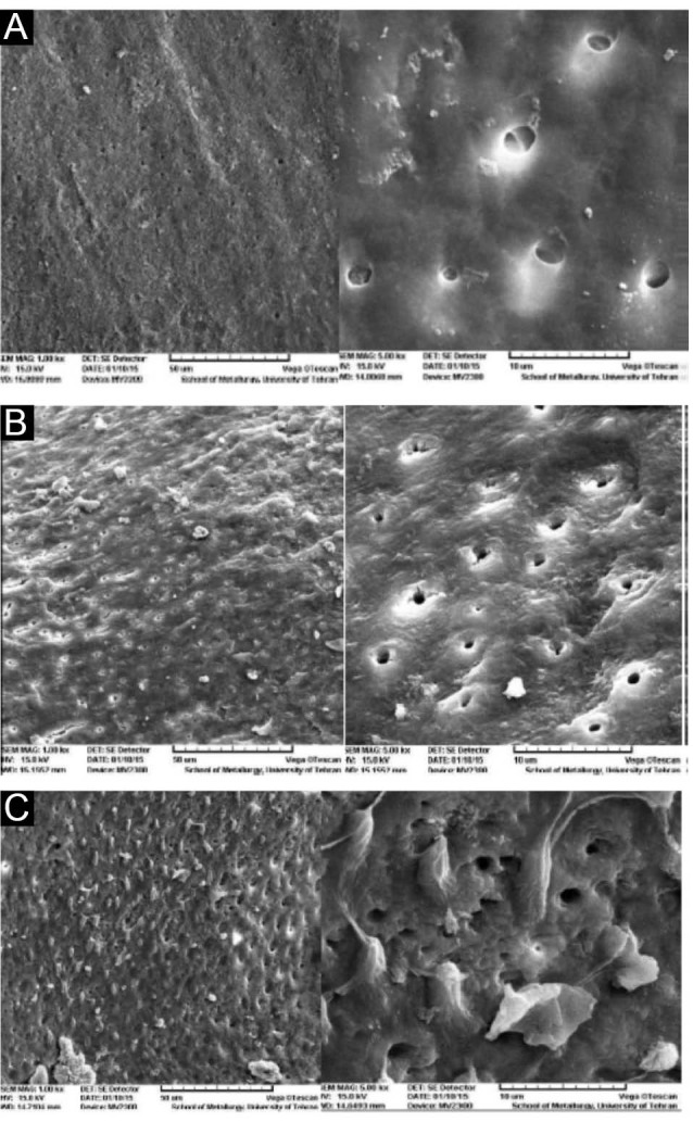
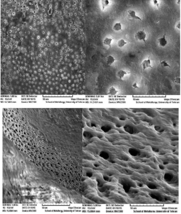
Introduction: The aim of this study was to evaluate the effect of various concentrations of NaOCl on shear bond strength of composite resin to dentin of primary teeth, prepared with laser and bur. Methods: In this in vitro study, 48 primary molars were sectioned at mesiodistal direction and were randomly divided into 6 groups; G1: bur, G2: bur + NaOCl 2.5%, G3: bur + NaOCl 5.25%, G4: laser, G5: laser + NaOCl 2.5%, G6: laser + NaOCl 5.25%. One-Step Plus adhesive was applied after phosphoric acid gel and NaOCl over the dentin surfaces for all groups, and composite resin cylinders were bonded to the samples. After thermocycling, shear bond strengths of composite resin to dentin were measured and statistical analyses were done by means of t test and analysis of variance (ANOVA). Results: The mean shear bond strength showed no significant difference between the groups prepared with bur (13.82 ± 3.49) and laser (14.18 ± 3.65) (P > 0.05). The mean difference of shear bond strength between three groups G1, G2 and G3 and between G4, G5 and G6 were not statistically significant (P > 0.05). Scanning electron microscopy (SEM) figures showed an irregular surface in laser groups and fairly complete removal of smear layer from the orifices of the dentinal tubules, in the group in which NaOCl was used. Conclusion: The application of different concentrations of NaOCl does not significantly improve the bond strength in dentin surfaces prepared with laser or bur.
Keywords: Electron scanning microscopy; Er:YAG Lasers; Hypochlorite; Primary teeth; Shear bond strength; Sodium.
Similar articles
-
Comparison between Shear Bond Strength of Er:YAG and Er,Cr:YSGG Lasers-Assisted Dentinal Adhesion of Self-Adhering Resin Composite: An Ex Vivo Study.Dent J (Basel). 2020 Jul 1;8(3):66. doi: 10.3390/dj8030066. Dent J (Basel). 2020. PMID: 32630313 Free PMC article.
-
Shear bond strength and ultrastructural interface analysis of different adhesive systems to Er:YAG laser-prepared dentin.Lasers Med Sci. 2015 Feb;30(2):769-78. doi: 10.1007/s10103-013-1424-0. Epub 2013 Aug 28. Lasers Med Sci. 2015. PMID: 23982720
-
Tensile bond strength of a flowable composite resin to ER:YAG-laser-treated dentin.Lasers Surg Med. 2005 Jun;36(5):351-5. doi: 10.1002/lsm.20176. Lasers Surg Med. 2005. PMID: 15825207
-
Deproteinization technique stabilizes the adhesion of the fiberglass post relined with resin composite to root canal.J Biomed Mater Res B Appl Biomater. 2012 Feb;100(2):577-83. doi: 10.1002/jbm.b.31946. Epub 2011 Nov 21. J Biomed Mater Res B Appl Biomater. 2012. PMID: 22102546 Review.
-
Effect of collagen crosslinkers on sodium hypochlorite treated dentin bond strength: a systematic review and meta-analysis.Front Bioeng Biotechnol. 2025 Apr 9;13:1547158. doi: 10.3389/fbioe.2025.1547158. eCollection 2025. Front Bioeng Biotechnol. 2025. PMID: 40271350 Free PMC article.
Cited by
-
Effect of Diode Laser and Fluoride Varnish on Microhardness of enamel: An In Vitro Study.J Lasers Med Sci. 2024 Dec 26;15:e69. doi: 10.34172/jlms.2024.69. eCollection 2024. J Lasers Med Sci. 2024. PMID: 39949473 Free PMC article.
-
Effect of Cavity Disinfectants on Adhesion to Primary Teeth-A Systematic Review.Int J Mol Sci. 2021 Apr 22;22(9):4398. doi: 10.3390/ijms22094398. Int J Mol Sci. 2021. PMID: 33922376 Free PMC article.
-
Removal of Composite Restoration from the Root Surface in the Cervical Region Using Er: YAG Laser and Drill-In Vitro Study.Materials (Basel). 2020 Jul 7;13(13):3027. doi: 10.3390/ma13133027. Materials (Basel). 2020. PMID: 32645864 Free PMC article.
-
The Influence of Erbium Laser Pretreatment on Dentin Shear Bond Strength and Bond Failure Modes: A Systematic Review and Network Meta-Analysis.J Adhes Dent. 2024 May 24;26:147-170. doi: 10.3290/j.jad.b5378611. J Adhes Dent. 2024. PMID: 38785223 Free PMC article.
-
Effect of Er:YAG laser cavity preparation on the bond strength of 2-hydroxyethyl methacrylate-free and 2-hydroxyethyl methacrylate-rich self-etch adhesive systems: An in vitro study.Dent Res J (Isfahan). 2019 Nov 12;16(6):389-397. eCollection 2019 Nov-Dec. Dent Res J (Isfahan). 2019. PMID: 31803385 Free PMC article.
References
-
- Hosain M, Yamada Y, Nakamura Y, Murakami Y, Tamaki Y, Matsumoto K. A study on surface roughnes and microleakage test in cavities prepared by Er:YAG laser irradiation and etched bur cavities. Lasers Med Sci. 2003;18:25–31. - PubMed
-
- Navarro RS, Gouw-Soares S, Cassoni A, Haypek P, Zezell DM, de Paula Eduardo C. The influence of erbium:yttrium–aluminum–garnet laser ablation with variable pulse width on morphology and microleakage of composite restorations. Lasers Med Sci. 2010;25(6):881–889. doi: 10.1007/s10103-009-0736-6. - DOI - PubMed
-
- Hibst R, Keller U. Experimental studies of the application of the Er:YAG laser on dental hard substances: I Measurement of the ablation rate. Lasers Surg Med. 1989;9:338–344. - PubMed
-
- Tokonabe H, Kouji R, Watanabe H, Nakamura Y, Matsumoto K. Morphological changes of human teeth with Er:YAG laser irradiation. J Clin Laser Med Surg. 1999;17:7–12. - PubMed
-
- Apel C, Schafer C, Gutknecht N. Demineralization of Er:YAG and Er,Cr:YSGG laser-prepared enamel cavities in vitro. Caries Res. 2003;37(1):34–37. - PubMed
LinkOut - more resources
Full Text Sources
Other Literature Sources
Research Materials
Miscellaneous