

Case Reports
doi: 10.1155/2017/4352474.
Epub 2017 Aug 24.
An Incidentally Detected Right Ventricular Pseudoaneurysm
Affiliations
- PMID: 28912975
- PMCID: PMC5587959
- DOI: 10.1155/2017/4352474
Item in Clipboard
Case Reports
An Incidentally Detected Right Ventricular Pseudoaneurysm
Case Rep Cardiol.
2017.
Abstract
Ventricular pseudoaneurysm is an uncommon, potentially fatal complication that has been associated with myocardial infarction, cardiac surgery, chest trauma, and infectious processes. Diagnosis can be challenging, as cases are rare and slowly progressing and typically lack identifiable features on clinical presentation. As a result, advanced imaging techniques have become the hallmark of identification. Ahead, we describe a patient who presents with acute decompensated heart failure and was incidentally discovered to have a large right ventricular pseudoaneurysm that developed following previous traumatic anterior rib fracture.
Figures
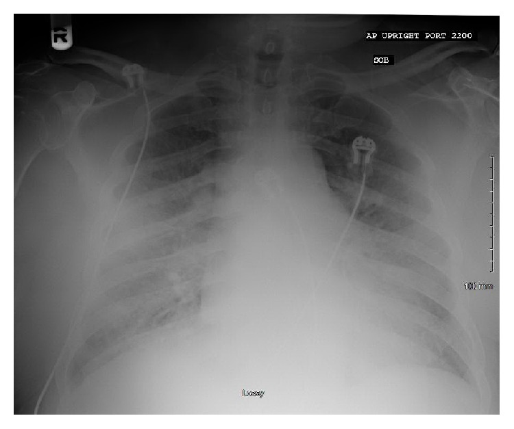
Portable chest X-ray. Minimal cardiomegaly and minimal interstitial pulmonary vascular congestion. Possible small right pleural effusion.
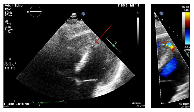
Transthoracic echocardiogram. Small, 5 mm ventricular septal defect in the distal inferoseptal/apical septum (red arrow).
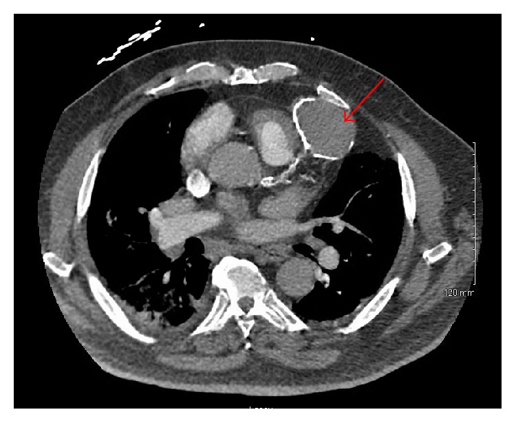
CT pulmonary angiogram. 6.4 × 4.3 cm peripherally calcified hyperattenuating lesion along the intraventricular groove highly concerning for a giant left anterior descending coronary artery aneurysm (red arrow).
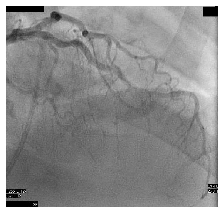
Left heart catheterization. Mild luminal irregularities of the left anterior descending artery and left circumflex arteries not requiring percutaneous intervention. No evidence of aneurysm.
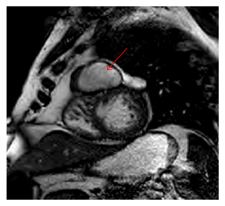
Cardiac MRI. 5.4 × 4.1 × 4.9 cm pseudoaneurysm arising from the mid-apical free wall of the right ventricle, with a 9 mm neck (red arrow). There is an adjacent muscular ventricular septal defect, which is likely related.
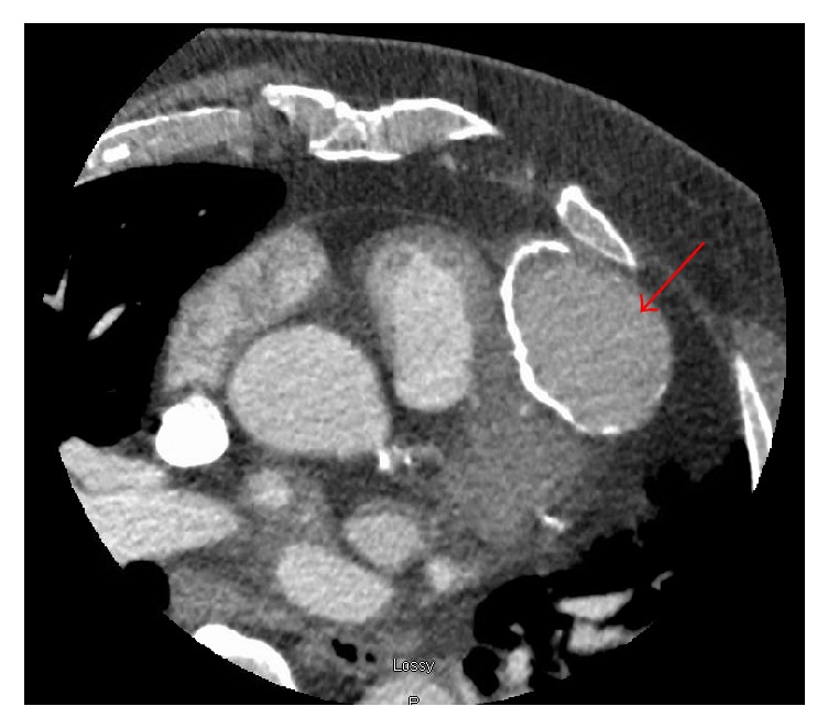
Cardiac CTA. Pseudoaneurysm arising superiorly from the mid-apical free wall of the right ventricle measuring up to 6.1 cm in maximal diameter with a 15 mm neck (red arrow).
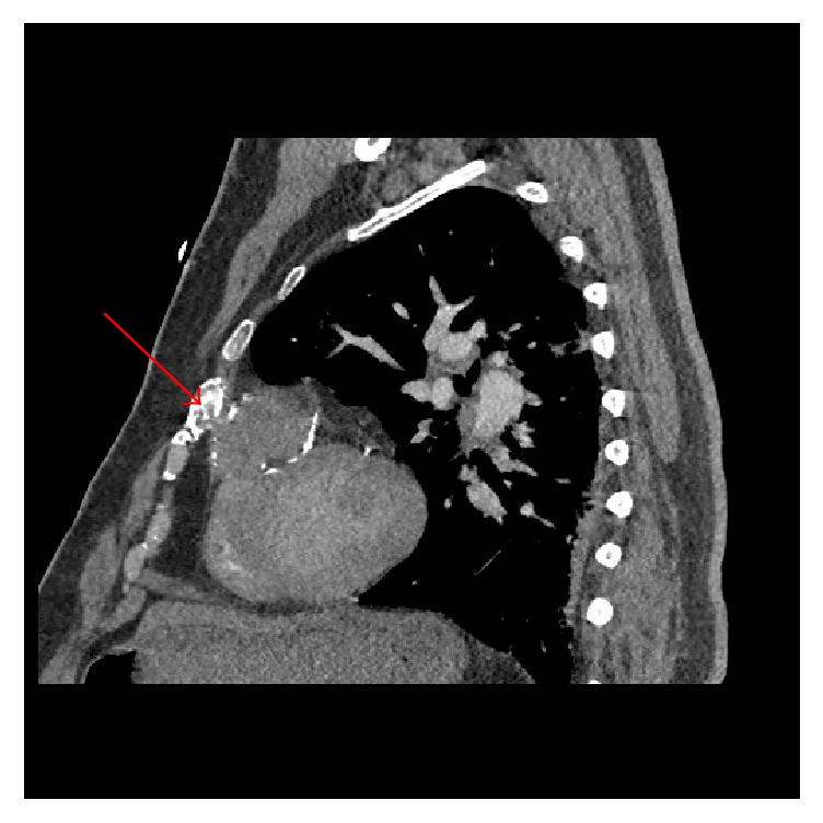
CT pulmonary angiogram. An old healed rib fracture anterior to the pseudoaneurysm (red arrow).
References
Publication types
LinkOut - more resources
Full Text Sources
Other Literature Sources
Research Materials
