

Case Reports
doi: 10.1016/j.rmcr.2017.08.019.
eCollection 2017.
Endobronchial hamartoma; a rare structural cause of chronic cough
Affiliations
- PMID: 28913162
- PMCID: PMC5587872
- DOI: 10.1016/j.rmcr.2017.08.019
Item in Clipboard
Case Reports
Endobronchial hamartoma; a rare structural cause of chronic cough
Respir Med Case Rep.
.
Abstract
Pulmonary hamartomas are rare benign tumors consisting of multiple mesenchymal cell lines like cartilage, bone and fat. We discuss an interesting case of a 53-year-old male patient, who was referred to our clinic for persistent cough. Chest X-ray revealed a left suprahilar density associated with plate like atelectasis, which on chest CT was found to be a densely calcified nodule, causing narrowing of the left upper lobe (LUL) bronchus with calcified bilateral hilar lymph nodes. A bronchoscopy revealed a smooth endobronchial mass with calcification, which was removed. Histopathology revealed pulmonary hamartoma.
Figures
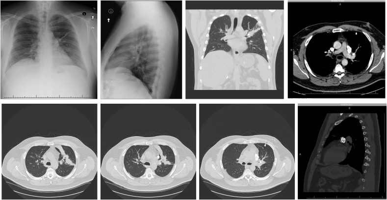
A: Dense calcified mass (arrow) overlying the left hilum associated with linear atelectasis extending laterally from the hilum and hyperinflation of the left upper lobe consistent with partial airway obstruction and peripheral air trapping. B: Chest X-ray Lateral View: Partial left upper lobe collapse and hilar calcifications. C: CT Chest Coronal View Without Contrast: Densely Calcified rounded mass causing LUL Bronchus narrowing and Calcified left hilar lymph nodes. D: CT Chest Axial View with contrast: Irregular Lobulated Calcified mass in LUL Bronchus causing plate like atelectasis with bilateral calcified hilar lymph nodes. E: CT Chest without contrast Axial View: Plate like LUL Partial Atelectasis due to LUL Bronchus narrowing. F: Caclified Lymphnodes in the Left Supra-hilar region. G: LUL Bronchus narrowing from a calcified endobronchial mass with surrounding non-calcified tissue. H: CT Chest Coronal View: Classic appearance of “popcorn calcification” in the left supra hilar region.

A: LUL Endobronchial Mass with smooth surface and inferolateral calcification. B: The anterior segment of the left upper lobe completely obstructed with scaring tissue and protrusion of the mass.
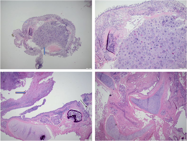
A: Benign lobulated mature cartilage ( ) covered by respiratory epithelium (
 ) (H&E biopsy specimen ×2). B: The benign Hyaline cartilage is hypercellular with slightly irregular surface (×10). C: Inflammatory polyp (
 ) and osseous metaplasia of peribronchial cartilage (
 ) and adjacent fat (
 ) (H&E resection specimen ×2). D: Disordered mass forming proliferation of mature cartilage, fibrovascular connective tissue, benign adipose tissue and slit like entrapped respiratory epithelium, consistent with Hamartoma. (H&E resection specimen ×2).
) covered by respiratory epithelium () (H&E biopsy specimen ×2). B: The benign Hyaline cartilage is hypercellular with slightly irregular surface (×10). C: Inflammatory polyp () and osseous metaplasia of peribronchial cartilage () and adjacent fat () (H&E resection specimen ×2). D: Disordered mass forming proliferation of mature cartilage, fibrovascular connective tissue, benign adipose tissue and slit like entrapped respiratory epithelium, consistent with Hamartoma. (H&E resection specimen ×2).References
Publication types
LinkOut - more resources
Full Text Sources
Other Literature Sources
