

Analysis of Facial Asymmetry
- PMID: 28913211
- PMCID: PMC5556787
- DOI: 10.7181/acfs.2015.16.1.1
Analysis of Facial Asymmetry
Abstract
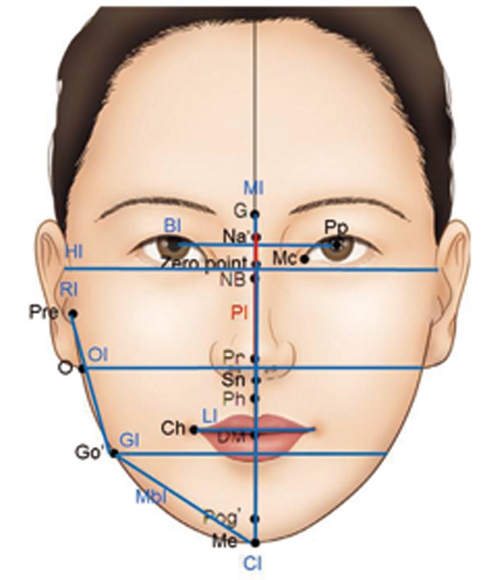
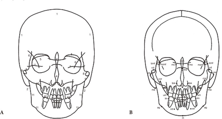
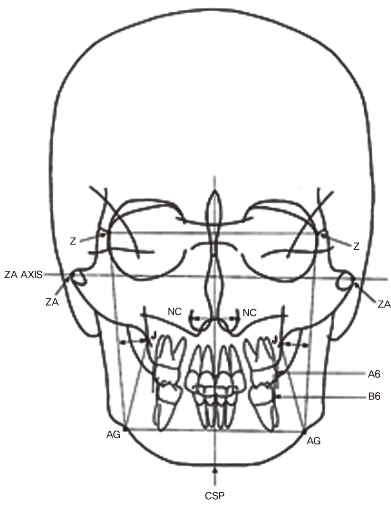
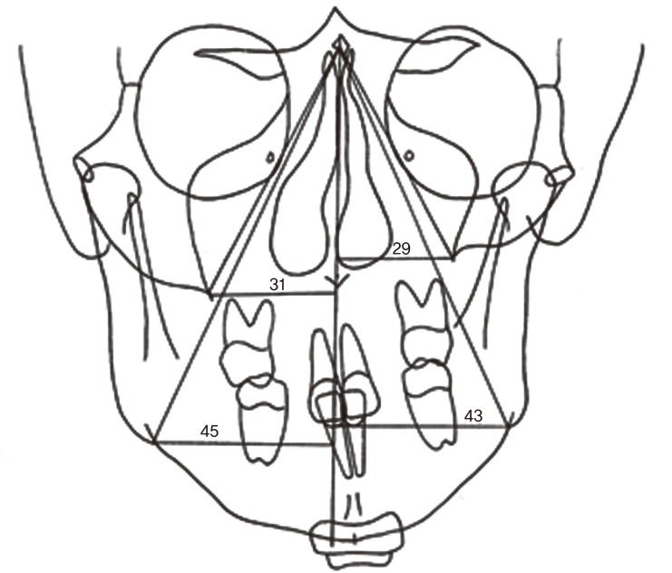
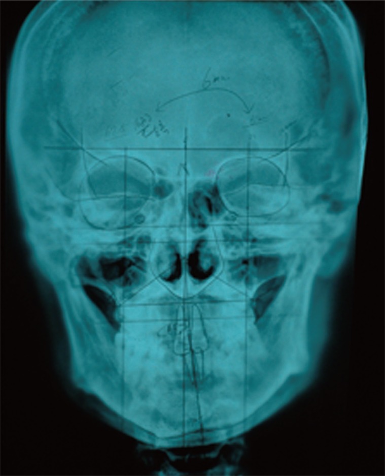
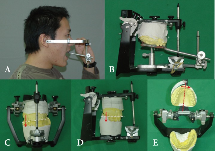
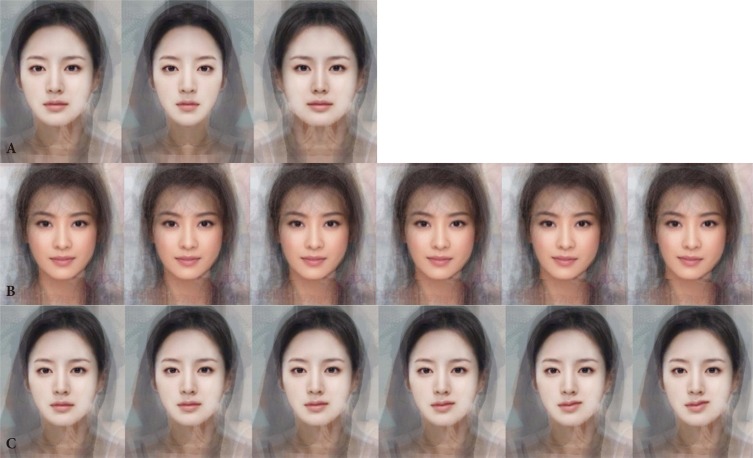
Facial symmetry is an important component of attractiveness. However, functional symmetry is favorable to aesthetic symmetry. In addition, fluctuating asymmetry is more natural and common, even if patients find such asymmetry to be noticeable. However, fluctuating asymmetry remains difficult to define. Several studies have shown that a certain level of asymmetry could generate an unfavorable image. A natural profile is favorable to perfect mirror-image profile, and images with canting and differences less than 3°-4° and 3-4 mm, respectively, are generally not recognized as asymmetry. In this study, a questionnaire survey among 434 medical students was used to evaluate photos of Asian women. The students preferred original images over mirror images. Facial asymmetry was noticed when the canting and difference were more than 3° and 3 mm, respectively. When a certain level of asymmetry is recognizable, correcting it can help to improve social life and human relationships. Prior to any operation, the anatomical component for noticeable asymmetry should be understood, which can be divided into hard tissues and soft tissue. For diagnosis, two-and three-dimensional (3D) photogrammetry and radiometry are used, including photography, laser scanner, cephalometry, and 3D computed tomography.
Keywords: Face; Facial asymmetry; Three dimensional image.
Conflict of interest statement
No potential conflict of interest relevant to this article was reported.
Figures
Similar articles
-
Reliability of a Method for Computing Facial Symmetry Plane and Degree of Asymmetry Based on 3D-data.J Orofac Orthop. 2007 Nov;68(6):477-90. doi: 10.1007/s00056-007-0652-y. J Orofac Orthop. 2007. PMID: 18034288 English, German.
-
Quantitative facial asymmetry: using three-dimensional photogrammetry to measure baseline facial surface symmetry.J Craniofac Surg. 2014 Jan;25(1):124-8. doi: 10.1097/SCS.0b013e3182a2e99d. J Craniofac Surg. 2014. PMID: 24406564
-
[Evaluation of therapeutic effect of virtual design for correcting facial asymmetry of skeletal Class Ⅲ deformity].Zhonghua Kou Qiang Yi Xue Za Zhi. 2016 Oct 9;51(10):594-599. doi: 10.3760/cma.j.issn.1002-0098.2016.10.005. Zhonghua Kou Qiang Yi Xue Za Zhi. 2016. PMID: 27719703 Chinese.
-
Quantification of facial asymmetry by 2D analysis - A comparison of recent approaches.J Craniomaxillofac Surg. 2014 Apr;42(3):265-71. doi: 10.1016/j.jcms.2013.07.033. Epub 2013 Sep 14. J Craniomaxillofac Surg. 2014. PMID: 24041610 Review.
-
Facial asymmetry: etiology, evaluation, and management.Chang Gung Med J. 2011 Jul-Aug;34(4):341-51. Chang Gung Med J. 2011. PMID: 21880188 Review.
Cited by
-
Analysis of hard tissue facial symmetry after unilateral mandibular reconstruction.Maxillofac Plast Reconstr Surg. 2021 Jun 1;43(1):15. doi: 10.1186/s40902-021-00299-2. Maxillofac Plast Reconstr Surg. 2021. PMID: 34059964 Free PMC article.
-
Development of a Cranial Suture Traction Therapy Program for Facial Asymmetry Correction Using the New Delphi Technique.Medicina (Kaunas). 2022 Jun 29;58(7):869. doi: 10.3390/medicina58070869. Medicina (Kaunas). 2022. PMID: 35888588 Free PMC article.
-
[Diagnostic and treatment alternatives for the correction of facial asymmetries: a literature review].Rev Cient Odontol (Lima). 2022 Mar 30;10(1):e098. doi: 10.21142/2523-2754-1001-2022-098. eCollection 2022 Jan-Mar. Rev Cient Odontol (Lima). 2022. PMID: 38389908 Free PMC article. Review. Spanish.
-
Is there a relationship between maxillary canine impaction and ocular asymmetry.J Orofac Orthop. 2019 Sep;80(5):236-241. doi: 10.1007/s00056-019-00189-3. Epub 2019 Aug 7. J Orofac Orthop. 2019. PMID: 31392346 English.
-
Exploring craniofacial fluctuating asymmetry in a South African sample.J Anat. 2025 Aug;247(2):314-334. doi: 10.1111/joa.14256. Epub 2025 Apr 23. J Anat. 2025. PMID: 40268871 Free PMC article.
References
-
- Chew MT. Spectrum and management of dentofacial deformities in a multiethnic Asian population. Angle Orthod. 2006;76:806–809. - PubMed
-
- Song WC, Koh KS, Kim SH, Hu KS, Kim HJ, Park JC, Choi BY. Horizontal angular asymmetry of the face in korean young adults with reference to the eye and mouth. J Oral Maxillofac Surg. 2007;65:2164–2168. - PubMed
-
- Haraguchi S, Takada K, Yasuda Y. Facial asymmetry in subjects with skeletal Class III deformity. Angle Orthod. 2002;72:28–35. - PubMed
-
- Giovanoli P, Tzou CH, Ploner M, Frey M. Three-dimensional videoanalysis of facial movements in healthy volunteers. Br J Plast Surg. 2003;56:644–652. - PubMed
-
- Bishara SE, Burkey PS, Kharouf JG. Dental and facial asymmetries: a review. Angle Orthod. 1994;64:89–98. - PubMed
Publication types
LinkOut - more resources
Full Text Sources
Other Literature Sources
