

Schwannoma of the Orbit
- PMID: 28913225
- PMCID: PMC5556852
- DOI: 10.7181/acfs.2015.16.2.67
Schwannoma of the Orbit
Abstract
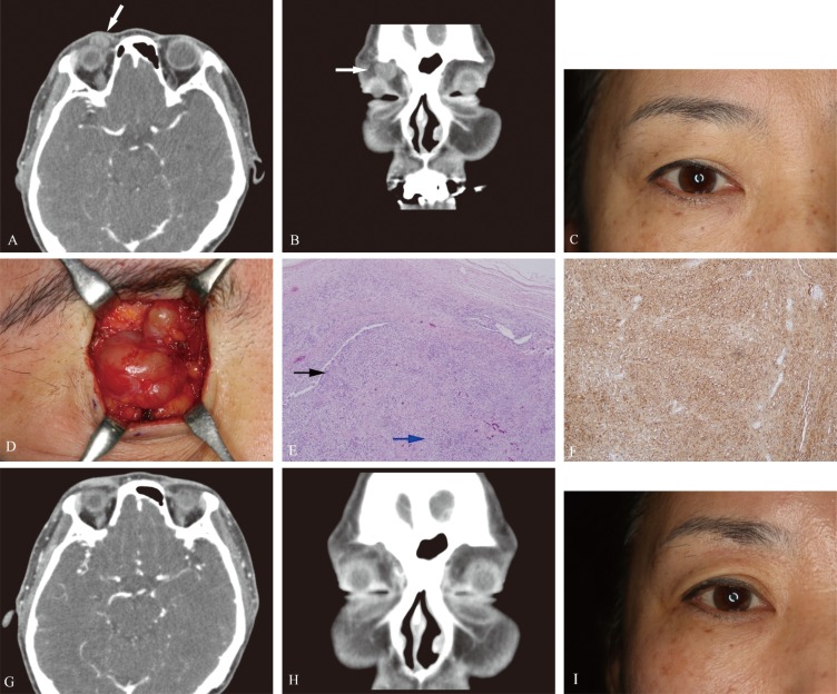
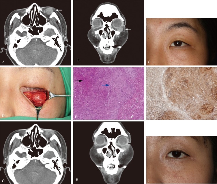
Background: A schwannoma is a benign, slow-growing peripheral nerve sheath tumor that originates from Schwann cells. Orbital schwannomas are rare, accounting for only 1% of all orbital neoplasms. In this study, we retrospectively review orbital schwannomas and characterize clinical, radiologic, and histologic features of this rare entity.
Methods: A retrospective review was performed to identify patients with histologically confirmed orbital schwannoma, among a list of 437 patients who had visited our hospital with soft tissue masses within the orbit as the primary presentation between 2010 and 2014. Patient charts and medical records were reviewed for demographic information, relevant medical and family history, physical examination findings relating to ocular and extraocular sensorimotor function, operative details, postoperative complications, pathologic report, and recurrence.
Results: Five patients (5/437, 1.1%) were identified as having histologically confirmed orbital schwannoma and underwent complete excision. Both computed tomography (CT) and magnetic resonance imaging (MRI) studies were not consistent in predicting histologic diagnosis. There were no complications, and none of the patients experienced significant scar formation. In two cases, patients exhibited a mild postoperative numbness of the forehead, but the patients demonstrated full recovery of sensation within 3 months after the operation. None of the five patients have experienced recurrence.
Conclusion: Orbital schwannomas are relatively rare tumors. Preoperative diagnosis is difficult because of its variable presentation and location. Appropriate early assessment of orbital tumors by CT or MRI and prompt management is warranted to prevent the development of severe complications. Therefore, orbital schwannomas should be considered in the differential diagnosis of slow-growing orbital masses.
Keywords: Neurilemmoma; Orbital neoplasm; Schwann cells; Surgery.
Conflict of interest statement
No potential conflict of interest relevant to this article was reported.
Figures
Similar articles
-
Orbital Schwannoma.J Neurol Surg B Skull Base. 2020 Aug;81(4):376-380. doi: 10.1055/s-0040-1713935. Epub 2020 Aug 24. J Neurol Surg B Skull Base. 2020. PMID: 33072479 Free PMC article.
-
Rare retro-orbital intraconal occurrence of benign schwannoma - a case report.J Clin Diagn Res. 2013 Dec;7(12):2964-5. doi: 10.7860/JCDR/2013/6827.3810. Epub 2013 Dec 15. J Clin Diagn Res. 2013. PMID: 24551692 Free PMC article.
-
Orbital schwannoma and neurofibroma: role of imaging.Neuroimaging Clin N Am. 2005 Feb;15(1):159-74. doi: 10.1016/j.nic.2005.02.004. Neuroimaging Clin N Am. 2005. PMID: 15927866 Review.
-
Schwannoma of the orbit.Ophthalmologica. 1984;188(2):118-27. doi: 10.1159/000309352. Ophthalmologica. 1984. PMID: 6709305
-
Pediatric intramedullary schwannoma with syringomyelia: a case report and literature review.BMC Pediatr. 2018 Nov 28;18(1):374. doi: 10.1186/s12887-018-1341-2. BMC Pediatr. 2018. PMID: 30486806 Free PMC article. Review.
Cited by
-
Orbital Tumors-Clinical, Radiologic and Histopathologic Correlation.Diagnostics (Basel). 2022 Sep 30;12(10):2376. doi: 10.3390/diagnostics12102376. Diagnostics (Basel). 2022. PMID: 36292065 Free PMC article. Review.
-
Imaging characteristics of orbital peripheral nerve sheath tumors: Analysis of 34 cases.World J Clin Cases. 2022 Jul 26;10(21):7356-7364. doi: 10.12998/wjcc.v10.i21.7356. World J Clin Cases. 2022. PMID: 36158022 Free PMC article.
-
Primary recurrent orbital schwannoma treated with surgical excision and Mitomycin-C.Am J Ophthalmol Case Rep. 2020 Jun 15;19:100784. doi: 10.1016/j.ajoc.2020.100784. eCollection 2020 Sep. Am J Ophthalmol Case Rep. 2020. PMID: 32613139 Free PMC article.
-
Pediatric dumbbell-shaped orbital schwannoma with extension to the cranial cavity: A case report and literature review.Front Neurol. 2023 Jan 10;13:1071632. doi: 10.3389/fneur.2022.1071632. eCollection 2022. Front Neurol. 2023. PMID: 36703626 Free PMC article.
-
Orbital Schwannoma.J Neurol Surg B Skull Base. 2020 Aug;81(4):376-380. doi: 10.1055/s-0040-1713935. Epub 2020 Aug 24. J Neurol Surg B Skull Base. 2020. PMID: 33072479 Free PMC article.
References
-
- Mora-Rios LE, Rios Y, Flores-Estrada JJ, Rodriguez-Reyes AA. Infraorbital schwannoma: case report. Cir Cir. 2014;82:76–80. - PubMed
-
- Volpe NJ, Gausas RE. Optic nerve and orbital tumors. Neurosurg Clin N Am. 1999;10:699–715. - PubMed
-
- Lam DS, Ng JS, To KF, Abdulah V, Liew CT, Tso MO. Cystic schwannoma of the orbit. Eye (Lond) 1997;11(Pt 6):798–800. - PubMed
-
- Brucoli M, Giarda M, Arcuri F, Benech A. A benign isolated schwannoma of the orbit. J Craniofac Surg. 2011;22:2372–2374. - PubMed
LinkOut - more resources
Full Text Sources
Other Literature Sources
