

Trichilemmal Carcinoma from Proliferating Trichilemmal Cyst on the Posterior Neck
- PMID: 28913304
- PMCID: PMC5556745
- DOI: 10.7181/acfs.2017.18.1.50
Trichilemmal Carcinoma from Proliferating Trichilemmal Cyst on the Posterior Neck
Abstract
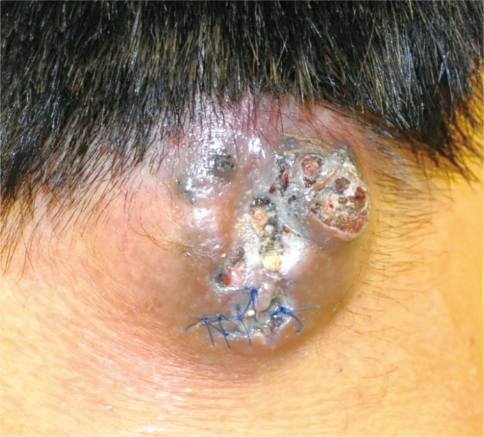
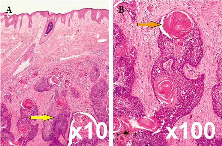
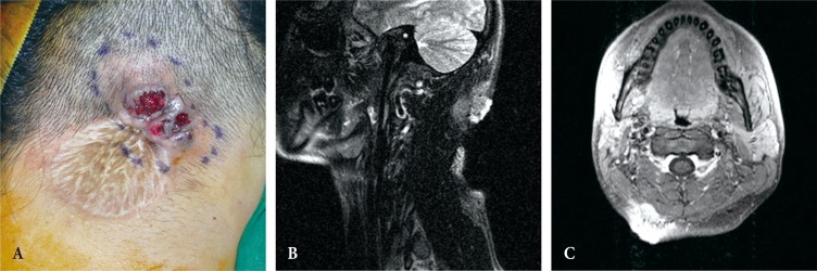
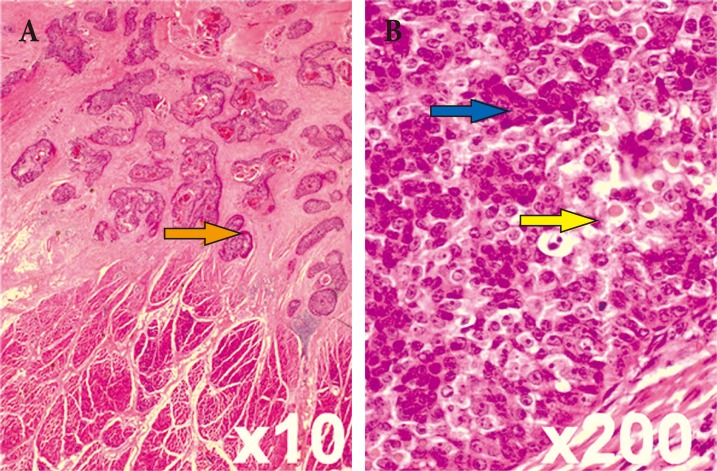
Trichilemmal cysts are common fluid-filled growths that arise from the isthmus of the hair follicle. They can form rapidly multiplying trichilemmal tumors-, also called proliferating trichilemmal cysts, which are typically benign. Rarely, proliferating trichilemmal cysts can become cancerous. Here we report the case of a patient who experienced this series of changes. The 27-year-old male patient had been observed to have a 1×1 cm cyst 7 years ago. Eight months prior to presentation at our institution, incision and drainage was performed at his local clinic. However, the size of the mass had gradually increased. At our clinic, he presented with a 5×4 cm hard mass that had recurred on the posterior side of his neck. The tumor was removed without safety margin, and the skin defect was covered with a split-thickness skin graft. The pathologic diagnosis was a benign proliferating trichilemmal cyst. The mass recurred after 4months, at which point, a wide excision (1.3-cm safety margin) and split-thickness skin graft were performed. The biopsy revealed a trichilemmal carcinoma arising from a proliferating trichilemmal cyst. This clinical experience suggests that clinicians should consider the possibility of malignant changes when diagnosing and treating trichilemmal cysts.
Keywords: Epidermal cyst; Hair disease; Skin appendage; Skin neoplasms.
Conflict of interest statement
No potential conflict of interest relevant to this article was reported.
Figures
References
-
- Brownstein MH, Arluk DJ. Proliferating trichilemmal cyst: a simulant of squamous cell carcinoma. Cancer. 1981;48:1207–1214. - PubMed
-
- Saida T, Oohara K, Hori Y, Tsuchiya S. Development of a malignant proliferating trichilemmal cyst in a patient with multiple trichilemmal cysts. Dermatologica. 1983;166:203–208. - PubMed
-
- Markal N, Kurtay A, Velidedeoglu H, Hucumenoglu S. Malignant transformation of a giant proliferating trichilemmal tumor of the scalp: patient report and literature review. Ann Plast Surg. 1998;41:314–316. - PubMed
-
- Yamaguchi J, Irimajiri T, Ohara K. Proliferating trichilemmal cyst arising in the arm of a young woman. Dermatology. 1994;189:90–92. - PubMed
-
- Casas JG, Woscoff A. Giant pilar tumor of the scalp. Arch Dermatol. 1980;116:1395. - PubMed
LinkOut - more resources
Full Text Sources
Other Literature Sources
