

Silicone Implant Sandwiched between Intact Nasal Bones with Fractured Nasal Bone Segments
- PMID: 28913306
- PMCID: PMC5556747
- DOI: 10.7181/acfs.2017.18.1.59
Silicone Implant Sandwiched between Intact Nasal Bones with Fractured Nasal Bone Segments
Abstract
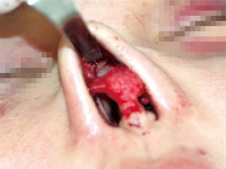
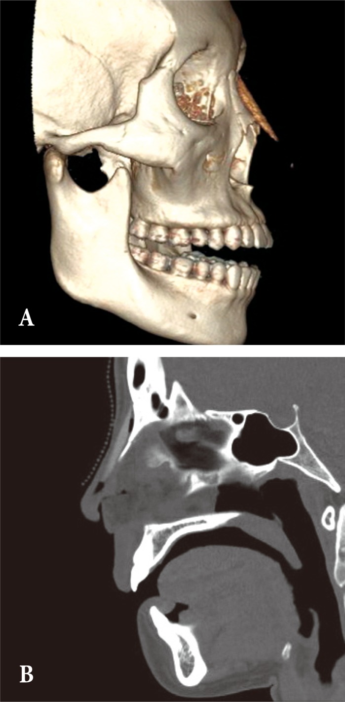
As the number of people who have undergone augmentation rhinoplasty has increased recently, nasal fractures are becoming more common after rhinoplasty. A silicone implant can affect the nasal fracture pattern, but there is no significant difference in treatment methods commonly. A 28-year-old female who had undergone augmentation visited our clinic with a nasal fracture. Computed tomography revealed that the silicone implant was sandwiched between the intact nasal bones with fractured bone fragments. In this case, open reduction was inevitable and a new silicone implant was inserted after reduction. Migration of the silicone implant beneath the nasal bone is a very rare phenomenon, but its accurate prevention and diagnosis is important because a closed reduction is impossible.
Keywords: Nasal bone; Rhinoplasty; Silicones.
Conflict of interest statement
No potential conflict of interest relevant to this article was reported.
Figures

Similar articles
-
Clinical Analysis of Nasal Bone Fracture in Patients Who Have Previously Undergone Dorsal Augmentation Using Silicone Implants: A Pilot Study.Aesthetic Plast Surg. 2019 Dec;43(6):1607-1614. doi: 10.1007/s00266-019-01410-9. Epub 2019 Jun 6. Aesthetic Plast Surg. 2019. PMID: 31172268
-
Postoperative Satisfaction in Nasal Bone Fracture Patients Who Had Rhinoplasty.J Craniofac Surg. 2016 Oct;27(7):1707-1710. doi: 10.1097/SCS.0000000000002928. J Craniofac Surg. 2016. PMID: 27748725
-
Spontaneous Fracture of a Silicone Implant: A Delayed Complication of Rhinoplasty.Ear Nose Throat J. 2023 Sep 25:1455613231200829. doi: 10.1177/01455613231200829. Online ahead of print. Ear Nose Throat J. 2023. PMID: 37743756
-
Unrecognized Fracture of a Silicone Implant During Closed Rhinoplasty.J Craniofac Surg. 2022 Sep 1;33(6):e552-e553. doi: 10.1097/SCS.0000000000008422. Epub 2021 Dec 14. J Craniofac Surg. 2022. PMID: 34907948
-
New nasal fracture classification for patients with silicone implants.J Plast Surg Hand Surg. 2013 Oct;47(5):363-7. doi: 10.3109/2000656X.2013.769442. Epub 2013 May 28. J Plast Surg Hand Surg. 2013. PMID: 23710785
References
-
- Chen CT, Hu TL, Lai JB, Chen YC, Chen YR. Reconstruction of traumatic nasal deformity in Orientals. J Plast Reconstr Aesthet Surg. 2010;63:257–264. - PubMed
-
- Jeon SP, Kang SJ, Kim JW, Kim YH, Sun H. New nasal fracture classification for patients with silicone implants. J Plast Surg Hand Surg. 2013;47:363–367. - PubMed
-
- Jung DH, Kim BR, Choi JY, Rho YS, Park HJ, Han WW. Gross and pathologic analysis of long-term silicone implants inserted into the human body for augmentation rhinoplasty: 221 revision cases. Plast Reconstr Surg. 2007;120:1997–2003. - PubMed
-
- Lin HT, Day YJ, Sum DC, Liu FC, Liou JT. Displaced nasal silicone implant: an unusual cause of nasotracheal tube obstruction. J Clin Anesth. 2013;25:344–345. - PubMed
-
- Hwang K, You SH, Kim SG, Lee SI. Analysis of nasal bone fractures; a six-year study of 503 patients. J Craniofac Surg. 2006;17:261–264. - PubMed
LinkOut - more resources
Full Text Sources
Other Literature Sources
Research Materials
