Review
. 2017:2017:4367019.
doi: 10.1155/2017/4367019. Epub 2017 Aug 23.

New Roles of the Primary Cilium in Autophagy

Affiliations
Review

New Roles of the Primary Cilium in Autophagy

Yenniffer Ávalos et al. Biomed Res Int. 2017.

Abstract

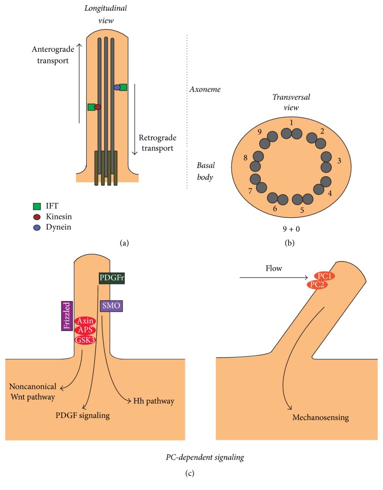
The primary cilium is a nonmotile organelle that emanates from the surface of multiple cell types and receives signals from the environment to regulate intracellular signaling pathways. The presence of cilia, as well as their length, is important for proper cell function; shortened, elongated, or absent cilia are associated with pathological conditions. Interestingly, it has recently been shown that the molecular machinery involved in autophagy, the process of recycling of intracellular material to maintain cellular and tissue homeostasis, participates in ciliogenesis. Cilium-dependent signaling is necessary for autophagosome formation and, conversely, autophagy regulates both ciliogenesis and cilium length by degrading specific ciliary proteins. Here, we will discuss the relationship that exists between the two processes at the cellular and molecular level, highlighting what is known about the effects of ciliary dysfunction in the control of energy homeostasis in some ciliopathies.


Figures

Figure 1
Figure 1
Primary cilium: structure and function. (a) Longitudinal representation of the primary cilium shows an axoneme center formed by highly stable acetylated microtubules that arise from the basal body parked near the nucleus. The components that are transported to the primary cilium (anterograde transport) rely on intraflagellar transport (IFT) proteins attached to Kinesins motor proteins. Conversely, transport from the primary cilium (retrograde transport) depends on different IFT proteins and dynein motor proteins. (b) Transversal representation of the primary cilium shows a 9 + 0 microtubule array, explaining the nonmotile behavior of the primary cilium. (c) Signaling of the primary cilium. On the left, several receptors present at the primary cilium such as platelet-derived growth factor receptor (PDGFR) for activation of the ERK pathway and Patched-1 (PTCH1) activated by Hh ligand for smoothened (SMO) translocation to the ciliary tip and activation of the transcription factor glioma (GLI). On the right, flow activates mechanosensation pathways by activation of the PC1/PC2 calcium channel and noncanonical activation of the Wnt pathway by increasing the expression of inversin in a calcium dependent mechanism, which participates in the degradation of APC and accumulation of β-catenin.
Figure 2
Figure 2
Overview of the autophagic pathway. Autophagy can be divided into five stages: initiation, nucleation, elongation, fusion, and degradation. “Initiation” refers to the activation of the ULK1 (ATG1) complex, which can be achieved by increased activity of AMP-dependent kinase (AMPK) and/or inhibition of the mechanistic target of rapamycin complex 1 (mTORC1). The “nucleation” indicates the process of recruitment of proteins of the autophagic machinery at the autophagosome formation sites, which are required for the formation of the new autophagosome, which occurs during the “elongation” stage. Then the complete autophagosome is loaded with the material that needs to be removed from the cell and degraded. The autophagosome fuses with a lysosome, thus activating the lysosomal acidic hydrolases, which degrade the cargo sequestered in the autophagic vesicle. See the main text for additional details.
Figure 3
Figure 3
Autophagy and primary cilium crosstalk. Summary of the published studies indicating the existence of a bidirectional crosstalk between ciliogenesis and the primary cilium. (a-b) Pampliega et al. show that proteins of the autophagic machinery localize at the primary cilium. In basal conditions, autophagy degrades the ciliary protein intraflagellar transport 20 (IFT20), inhibiting ciliary growth. In addition, when autophagy is induced by serum starvation, ciliogenesis is promoted by a cilium-dependent activation of the Hedgehog pathway. (c) Tang et al. show that ciliogenesis is induced in conditions of serum starvation through autophagy-mediated degradation of the protein oral-facial-digital syndrome 1 (OFD1). (d) Wang et al. work indicates that autophagy and primary cilium reciprocally affect each other: inhibition of ciliogenesis is associated with the activation of mechanistic target of rapamycin (mTOR) and consequently with the inhibition of autophagy. On the other side, a condition of impaired autophagy stimulates the proteasome, which inhibits ciliogenesis. See the main text for additional details.
Figure 4
Figure 4
Effects of ciliary dysfunction. Depletion or shortening of PC alters the production of hypothalamic neuropeptides increasing food intake and therefore fat mass. In addition, animals showing ciliary dysfunction are leptin and insulin resistant and intolerant to glucose. In conclusion, depletion or shortening of PC drives obesity and obesity associated diseases. The mechanisms that drive these effects are far from being understood and we propose that this occurs because of an impairment in autophagy. See the main text for additional details.

Similar articles

Cited by

References

    1. Malicki J. J., Johnson C. A. The Cilium: Cellular antenna and central processing unit. Trends in Cell Biology. 2017;27(2):126–140. doi: 10.1016/j.tcb.2016.08.002. - DOI - PMC - PubMed
    1. Davenport J. R., Watts A. J., Roper V. C., et al. Disruption of Intraflagellar Transport in Adult Mice Leads to Obesity and Slow-Onset Cystic Kidney Disease. Current Biology. 2007;17(18):1586–1594. doi: 10.1016/j.cub.2007.08.034. - DOI - PMC - PubMed
    1. Oh E. C., Vasanth S., Katsanis N. Metabolic regulation and energy homeostasis through the primary Cilium. Cell Metabolism. 2015;21(1):21–31. doi: 10.1016/j.cmet.2014.11.019. - DOI - PMC - PubMed
    1. Marino G., Madeo F., Kroemer G. Autophagy for tissue homeostasis and neuroprotection. Curr Opin Cell Biol. 2011;23(2):198–206. doi: 10.1016/j.ceb.2010.10.001. - DOI - PubMed
    1. Pampliega O., Orhon I., Patel B., et al. Functional interaction between autophagy and ciliogenesis. Nature. 2013;502(7470):194–200. doi: 10.1038/nature12639. - DOI - PMC - PubMed

LinkOut - more resources