

Keratoconus diagnosis using Corvis ST measured biomechanical parameters
- PMID: 28913507
- PMCID: PMC5587249
- DOI: 10.1016/j.joco.2017.05.002
Keratoconus diagnosis using Corvis ST measured biomechanical parameters
Abstract
Purpose: To assess the diagnostic power of the Corneal Visualization Scheimpflug Technology (Corvis ST) provided corneal biomechanical parameters in keratoconic corneas.
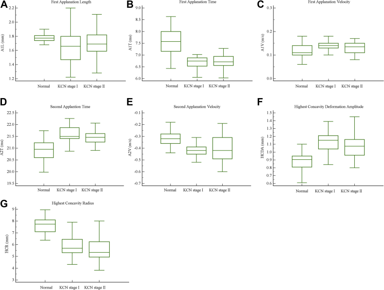
Methods: The following biomechanical parameters of 48 keratoconic eyes were compared with the corresponding ones in 50 normal eyes: time of the first applanation and time from start to the second applanation [applanation-1 time (A1T) and applanation-2 time (A2T)], time of the highest corneal displacement [highest concavity time (HCT)], magnitude of the displacement [highest concavity deformation amplitude (HCDA)], the length of the flattened segment in the applanations [first applanation length (A1L) and second applanation length (A2L)], velocity of corneal movement during applanations [applanation-1 velocity (A1V) and applanation-2 velocity (A2V)], distance between bending points of the cornea at the highest concavity [highest concavity peak distance (HCPD)], central concave curvature at the highest concavity [highest concavity radius (HCR)]. To assess the change of parameters by disease severity, the keratoconus group was divided into two subgroups, and their biomechanical parameters were compared with each other and with normal group. The parameters' predictive ability was assessed by receiver operating characteristic (ROC) curves. To control the effect of central corneal thickness (CCT) difference between the two groups, two subgroups with similar CCT were selected, and the analyses were repeated.
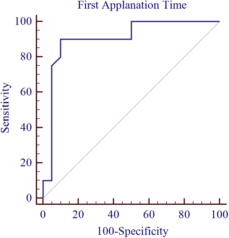
Results: Of the 10 parameters compared, the means of the 8 were significantly different between groups (P < 0.05). Means of the parameters did not show significant difference between keratoconus subgroups (P > 0.05). ROC curve analyses showed excellent distinguishing ability for A1T and HCR [area under the curve (AUC) > 0.9], and good distinguishing ability for A2T, A2V, and HCDA (0.9 > AUC > 0.7). A1T reading was able to correctly identify at least 93% of eyes with keratoconus (cut-off point 7.03). In two CCT matched subgroups, A1T showed an excellent distinguishing ability again.
Conclusions: The A1T seems a valuable parameter in the diagnosis of keratoconic eyes. It showed excellent diagnostic ability even when controlled for CCT. None of the parameters were reliable index for keratoconus staging.
Keywords: Biomechanics; Cornea; Corvis ST; Keratoconus.
Figures
Similar articles
-
Corneal biomechanical metrics of healthy Chinese adults using Corvis ST.Cont Lens Anterior Eye. 2017 Apr;40(2):97-103. doi: 10.1016/j.clae.2016.12.003. Epub 2016 Dec 10. Cont Lens Anterior Eye. 2017. PMID: 27964894
-
Biomechanical assessment of healthy and keratoconic corneas (with/without crosslinking) using dynamic ultrahigh-speed Scheimpflug technology and the relevance of the parameter (A1L-A2L).Br J Ophthalmol. 2019 Apr;103(4):558-564. doi: 10.1136/bjophthalmol-2017-311627. Epub 2018 Jun 5. Br J Ophthalmol. 2019. PMID: 29871966
-
[Examination and discriminant analysis of corneal biomechanics with CorVis ST in keratoconus and subclinical keratoconus].Beijing Da Xue Xue Bao Yi Xue Ban. 2019 Oct 18;51(5):881-886. doi: 10.19723/j.issn.1671-167X.2019.05.015. Beijing Da Xue Xue Bao Yi Xue Ban. 2019. PMID: 31624393 Free PMC article. Chinese.
-
Corneal Biomechanical Characteristics in Myopes and Emmetropes Measured by Corvis ST: A Meta-Analysis.Am J Ophthalmol. 2024 Aug;264:154-161. doi: 10.1016/j.ajo.2024.03.024. Epub 2024 Mar 29. Am J Ophthalmol. 2024. PMID: 38556185
-
Corneal Parameters in Healthy Subjects Assessed by Corvis ST.J Ophthalmic Vis Res. 2020 Feb 2;15(1):24-31. doi: 10.18502/jovr.v15i1.5936. eCollection 2020 Jan-Mar. J Ophthalmic Vis Res. 2020. PMID: 32095205 Free PMC article. Review.
Cited by
-
Corneal biomechanical changes in allergic conjunctivitis.Eye Vis (Lond). 2021 May 3;8(1):17. doi: 10.1186/s40662-021-00241-7. Eye Vis (Lond). 2021. PMID: 33934706 Free PMC article.
-
Diagnosis of Subclinical Keratoconus with a Combined Model of Biomechanical and Topographic Parameters.J Clin Med. 2021 Jun 22;10(13):2746. doi: 10.3390/jcm10132746. J Clin Med. 2021. PMID: 34206580 Free PMC article.
-
Keratectasia severity staging and progression assessment based on the biomechanical E-staging.Eye Vis (Lond). 2024 Jul 1;11(1):24. doi: 10.1186/s40662-024-00392-3. Eye Vis (Lond). 2024. PMID: 38946004 Free PMC article. Review.
-
Characterization of non-linear mechanical behavior of the cornea.Sci Rep. 2020 Jul 14;10(1):11549. doi: 10.1038/s41598-020-68391-7. Sci Rep. 2020. PMID: 32665558 Free PMC article.
-
Comparisons of corneal biomechanical and tomographic parameters among thin normal cornea, forme fruste keratoconus, and mild keratoconus.Eye Vis (Lond). 2021 Nov 16;8(1):44. doi: 10.1186/s40662-021-00266-y. Eye Vis (Lond). 2021. PMID: 34784958 Free PMC article.
References
-
- Vellara H.R., Patel D.V. Biomechanical properties of the keratoconic cornea: a review. Clin Exp Optom. 2015;98:31–38. - PubMed
-
- Schweitzer C., Roberts C.J., Mahmoud A.M., Colin J., Maurice-Tison S., Kerautret J. Screening of forme fruste keratoconus with the ocular response analyzer. Invest Ophthalmol Vis Sci. 2010;51:2403–2410. - PubMed
-
- O'Keefe M., Kirwan C. Laser epithelial keratomileusis in 2010 – a review. Clin Exp Ophthalmol. 2010;38:183–191. - PubMed
-
- Ali N.Q., Patel D.V., McGhee C.N. Biomechanical responses of healthy and keratoconic corneas measured using a noncontact Scheimpflug-based tonometer. Invest Ophthalmol Vis Sci. 2014;55:3651–3659. - PubMed
LinkOut - more resources
Full Text Sources
Other Literature Sources
