. 2017 Sep 14;112(6):62.
doi: 10.1007/s00395-017-0652-z.

Dual inhibition of cathepsin G and chymase reduces myocyte death and improves cardiac remodeling after myocardial ischemia reperfusion injury

Affiliations

Dual inhibition of cathepsin G and chymase reduces myocyte death and improves cardiac remodeling after myocardial ischemia reperfusion injury

Bahman Hooshdaran et al. Basic Res Cardiol. .

Abstract

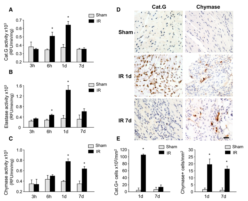
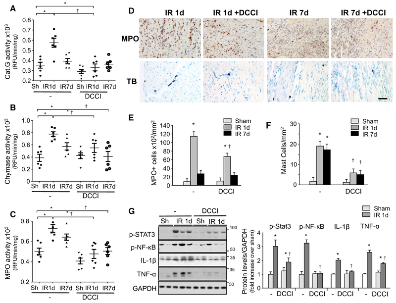
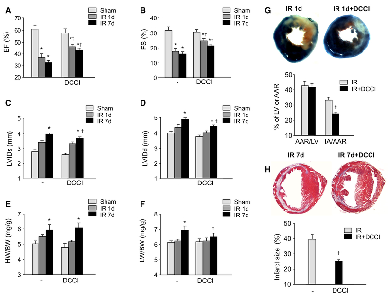
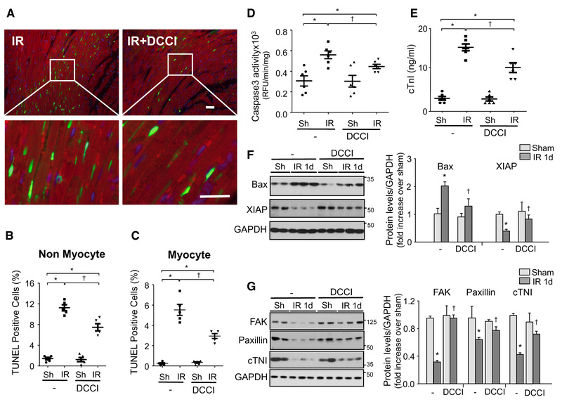
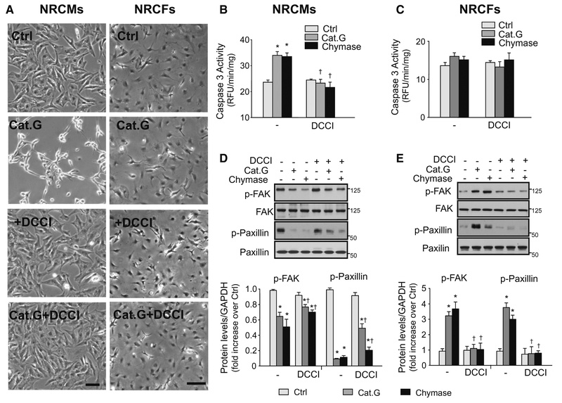
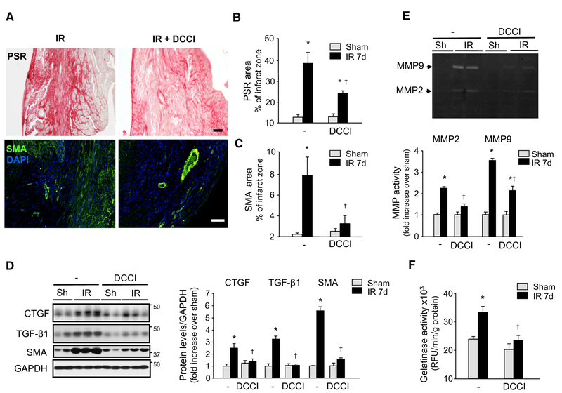
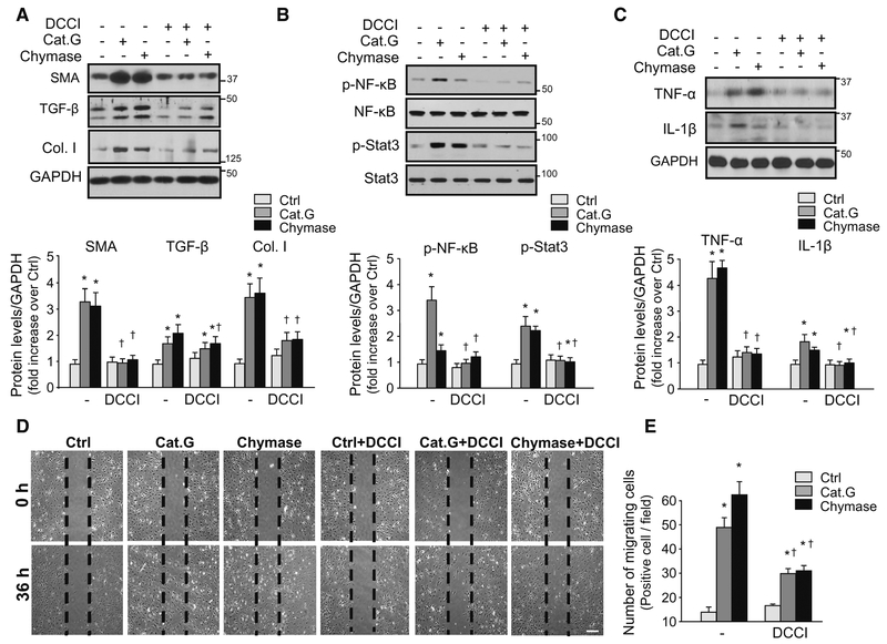
Early reperfusion of ischemic cardiac tissue increases inflammatory cell infiltration which contributes to cardiomyocyte death and loss of cardiac function, referred to as ischemia/reperfusion (IR) injury. Neutrophil- and mast cell-derived proteases, cathepsin G (Cat.G) and chymase, are released early after IR, but their function is complicated by potentially redundant actions and targets. This study investigated whether a dual inhibition of Cat.G and chymase influences cardiomyocyte injury and wound healing after experimental IR in mice. Treatment with a dual Cat.G and chymase inhibitor (DCCI) immediately after reperfusion blocked cardiac Cat.G and chymase activity induced after IR, which resulted in decreased immune response in the infarcted heart. Mice treated with DCCI had less myocardial collagen deposition and showed preserved ventricular function at 1 and 7 days post-IR compared with vehicle-treated mice. DCCI treatment also significantly attenuated focal adhesion (FA) complex disruption and myocyte degeneration after IR. Treatment of isolated cardiomyocytes with Cat.G or chymase significantly promoted FA signaling downregulation, myofibril degeneration and myocyte apoptosis. Conversely, treatment of cardiac fibroblasts with Cat.G or chymase induced FA signaling activation and increased their migration and differentiation to myofibroblasts. These opposite responses in cardiomyocytes and fibroblasts were blocked by treatment with DCCI. These findings show that Cat.G and chymase are key mediators of myocyte apoptosis and fibroblast migration and differentiation that play a role in adverse cardiac remodeling and function post-IR. Thus, dual targeting of neutrophil- and mast cell-derived proteases could be used as a novel therapeutic strategy to reduce post-IR inflammation and improve cardiac remodeling.

Keywords: Cardioprotection; Inflammation; Inflammatory serine proteases; Myocardial ischemia–reperfusion.


Conflict of interest statement

Conflict of interest The authors declare that they have no conflict of interest.

Figures

Fig. 1
Fig. 1
Inflammatory serine proteases are elevated after ischemia reperfusion (IR). The left anterior descending artery was ligated for 30 min to induce ischemia and the heart was subsequently reperfused for 3 h, 6 h, 1 or 7 days. Cathepsin G (Cat.G) (a), elastase (b) and chymase (c) activity levels in the hearts of sham and mice subjected to IR injury were assessed by substrate specific enzymatic activity assay. Results are expressed as relative fluorescence units (RFU)/min/mg protein (n = 6 for each group). d Representative Cat.G and chymase immunostaining of paraffin-embedded heart sections of sham or mice subjected to IR injury. Scale bar: 40 μm. e Quantification of Cat.G and chymase-positive cells (n = 5 for each group). Values are presented as mean ± SEM, *P < 0.05 vs. WT shams
Fig. 2
Fig. 2
DCCI treatment attenuates inflammation in the reperfused heart. Cathepsin G (Cat.G) (a), chymase (b) and myeloperoxidase (MPO) (c) activity in the infarcted LV as determined by enzymatic activity assay (n = 6 for each group). d Representative MPO or toluidine blue (TB) staining of paraffin-embedded heart sections from sham or mice subjected to ischemia reperfusion (IR). Scale bar: 40 μm. e, f Quantification of MPO- and mast cell-positive cells in mice treated with either vehicle or DCCI (n = 5 for each group). g Left: immunoblot analysis indicates a decrease in inflammatory signaling in the infarcted region of mice treated with DCCI compared to vehicle post-IR. Right: quantification of experiments represented as fold change compared to sham animals treated with vehicle (n = 5 for each group). GAPDH was included as a loading control. Values are presented as mean ± SEM, *P < 0.05 vs. shams, P < 0.05 vs. vehicle-treated IR
Fig. 3
Fig. 3
DCCI improves cardiac function post-IR. Echocardiography measurement of left ventricular (LV) ejection fraction (EF) (a), fractional shortening (FS) (b) and LV internal diameter (LVID) in systole (c) and diastole (d). Effects of DCCI treatment on IR-induced heart weight (HW) (e) or lung weight (LW) (f) to body weight (BW) ratio (n = 6 shams, n = 8 IR groups). Top: representative images of Evans blue and diphenyl tetrazolium chloride or Masson’s trichrome staining on transverse heart sections at day 1 (g) and 7 (h) post-IR, respectively. Bottom: quantification of the area at risk (AAR) and infarct area (IA) (n = 5 for each group). Values are presented as mean ± SEM, *P < 0.05 vs. sham, P < 0.05 vs. vehicle-treated IR
Fig. 4
Fig. 4
DCCI deletion offers cardioprotection after acute IR. Ischemia reperfusion (IR) was induced for 24 h. a LV tissue sections were assessed for apoptosis using TUNEL assay (green), tropomyosin (red), and DAPI (4′,6-diamidino-2-phenylindole) (blue) staining. Scale bar: 40 μm. The number of TUNEL-positive myocytes (b) and non-myocytes (c) in the reperfused area was expressed as a percentage of total nuclei detected by DAPI staining (n = 5 each group). d Quantification of caspase-3 activity in the LV using caspase-3 specific fluorogenic substrate (n = 6 each group). e Plasma levels of cardiac troponin-I (cTnI) as determined by ELISA assay in samples from vehicle- or DCCI-treated mice 24 h post-IR (n = 6 each group). Immunoblot analysis indicates a decrease in pro-apoptotic signaling (f) and focal adhesion signaling (g) in the infarcted region of mice treated with DCCI or vehicle post-IR. Left: representative immunoblots. Right: quantification of experiments represented as fold change compared to shams (n = 6 each group). GAPDH was included as a loading control. Values are presented as mean ± SEM, *P < 0.05 vs. shams, P < 0.05 vs. vehicle-treated
Fig. 5
Fig. 5
Dual effects of cathepsin G and chymase on focal adhesion signaling and cell detachment. Neonatal rat cardiomyocytes (a, b, d) and fibroblasts (a, c, e) were pretreated without or with DCCI (5 μM) prior to treatment with cathepsin G (Cat.G) or chymase for 8 h. a Phase-contrast photomicrographs (bar = 100 μm). b, c Caspase-3 activity was measured using fluorogenic substrate. d, e Left: representative immunoblots showing focal adhesion (FA) signaling in cardiomyocytes (d) and fibroblasts (f) treated with Cat.G or chymase for 10 min in the presence of DCCI or its vehicle. Right: quantification of experiments expressed as mean ± SEM from three separate cultures. *P < 0.05 vs. control, P < 0.05 vs. vehicle-treated cells
Fig. 6
Fig. 6
DCCI reduces fibrosis and improves cardiac remodeling after IR. Ischemia reperfusion (IR) was induced for 7 days. a Picro-sirus red staining and immunohistochemistry against smooth muscle α-actin (SMA) show reduced collagen and myofibroblast deposition in the injured myocardium of mice treated with DCCI compared to those treated with vehicle. Scale bar: 40 μm. Semi-quantitative analysis of collagen staining (b) and SMA immunoreactive myofibroblasts (c) expressed as a percentage of total infarct area. d Immunoblot analysis indicates a decrease in profibrotic signaling molecules in the infarcted region of mice treated with DCCI vs. the treatment with vehicle post-IR. Left: representative immunoblots. Right: quantification of experiments represented as fold change compared to WT animals (n = 6 for each group). GAPDH was included as a loading control. e In-gel zymography (top) and relative band density quantification (bottom) indicate a decrease in MMP-2 and −9 activity in the infarcted myocardium of mice treated with DCCI vs. treated with vehicle. f Gelatinase activity assay in animals subjected to IR and treated with either vehicle or DCCI (n = 5 for each group). Values are presented as mean ± SEM, *P < 0.05 vs. shams, P < 0.05 vs. vehicle-treated IR
Fig. 7
Fig. 7
Cathepsin G and chymase induce cardiac fibroblast migration and differentiation. ac Neonatal rat cardiac fibroblasts were pretreated with DCCI (5 μM) or vehicle for 15 min and then treated with cathepsin G (Cat.G, 0.02 U/ml) or chymase (100 nM) for 36 h and cell lysates were processed for immunoblot analysis. Top: representative immunoblots. Bottom: quantification of experiments represented as fold change compared to untreated control (Ctrl). GAPDH was included as a loading control. d Migration scratch assay was performed to assess the rate of migration of cardiac fibroblasts untreated or treated with Cat.G or chymase with or without DCCI pretreatment. e A significant increase in the migration rate was observed in Cat.G- and chymase-treated fibroblasts compared with controls which was attenuated by DCCI treatment. Scale bar: 200 μm. Results are representative of three independent experiments. Data are mean ± SEM; *P < 0.05 vs. control; P < 0.05 vs. vehicle-treated fibroblasts

References

    1. Beghdadi W, Madjene LC, Benhamou M, Charles N, Gautier G, Launay P, Blank U (2011) Mast cells as cellular sensors in inflammation and immunity. Front Immunol 2:37. doi:10.3389/fimmu.2011.00037 - DOI - PMC - PubMed
    1. Caughey GH (2016) Mast cell proteases as pharmacological targets. Eur J Pharmacol 778:44–55. doi:10.1016/j.ejphar.2015.04.045 - DOI - PMC - PubMed
    1. Ducharme A, Frantz S, Aikawa M, Rabkin E, Lindsey M, Rohde LE, Schoen FJ, Kelly RA, Werb Z, Libby P, Lee RT (2000) Targeted deletion of matrix metalloproteinase-9 attenuates left ventricular enlargement and collagen accumulation after experimental myocardial infarction. J Clin Investig 106:55–62. doi:10.1172/JCI8768 - DOI - PMC - PubMed
    1. Frangogiannis NG (2012) Regulation of the inflammatory response in cardiac repair. Circ Res 110:159–173. doi:10.1161/CIRCRESAHA.111.243162 - DOI - PMC - PubMed
    1. Fu L, Wei C-C, Powell PC, Bradley WE, Ahmad S, Ferrario CM, Collawn JF, Dell’Italia LJ (2016) Increased fibroblast chymase production mediates procollagen autophagic digestion in volume overload. J Mol Cell Cardiol 92:1–9. doi:10.1016/j.yjmcc.2016.01.019 - DOI - PMC - PubMed

Publication types

MeSH terms

LinkOut - more resources