

The mediating role of cortical thickness and gray matter volume on sleep slow-wave activity during adolescence
- PMID: 28913599
- PMCID: PMC5828920
- DOI: 10.1007/s00429-017-1509-9
The mediating role of cortical thickness and gray matter volume on sleep slow-wave activity during adolescence
Abstract
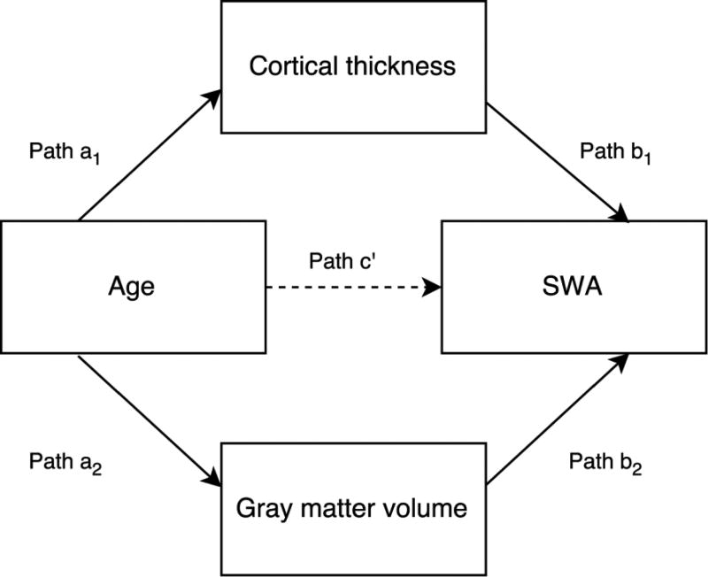
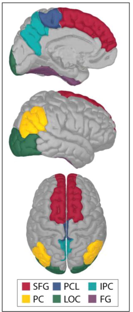
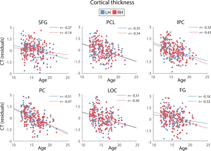
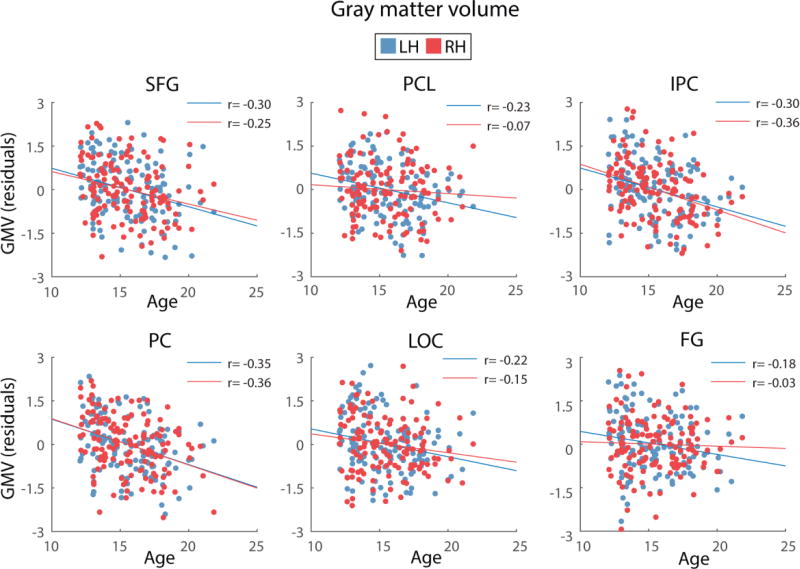
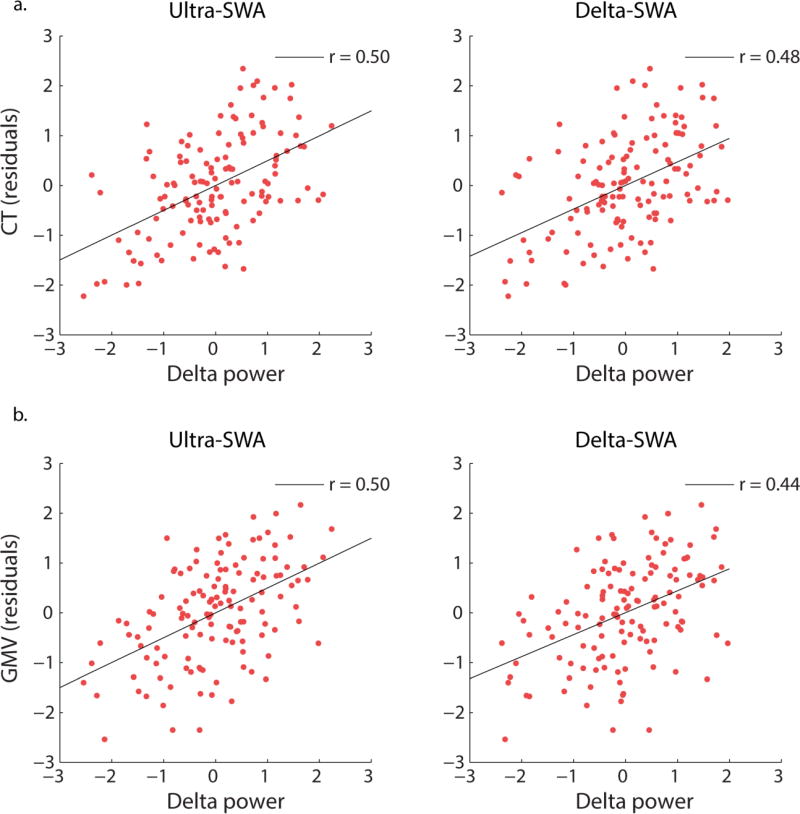
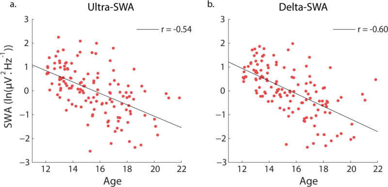
During the course of adolescence, reductions occur in cortical thickness and gray matter (GM) volume, along with a 65% reduction in slow-wave (delta) activity during sleep (SWA) but empirical data linking these structural brain and functional sleep differences, is lacking. Here, we investigated specifically whether age-related differences in cortical thickness and GM volume and cortical thickness accounted for the typical age-related difference in slow-wave (delta) activity (SWA) during sleep. 132 healthy participants (age 12-21 years) from the National Consortium on Alcohol and NeuroDevelopment in Adolescence study were included in this cross-sectional analysis of baseline polysomnographic, electroencephalographic, and magnetic resonance imaging data. By applying mediation models, we identified a large, direct effect of age on SWA in adolescents, which explained 45% of the variance in ultra-SWA (0.3-1 Hz) and 52% of the variance in delta-SWA (1 to <4 Hz), where SWA was lower in older adolescents, as has been reported previously. In addition, we provide evidence that the structure of several, predominantly frontal, and parietal brain regions, partially mediated this direct age effect, models including measures of brain structure explained an additional 3-9% of the variance in ultra-SWA and 4-5% of the variance in delta-SWA, with no differences between sexes. Replacing age with pubertal status in models produced similar results. As reductions in GM volume and cortical thickness likely indicate synaptic pruning and myelination, these results suggest that diminished SWA in older, more mature adolescents may largely be driven by such processes within a number of frontal and parietal brain regions.
Keywords: Adolescence; Cortical development; Sleep; Slow-wave activity.
Conflict of interest statement
Conflict of interest: The authors declare no conflicts of interest
Figures
Similar articles
-
EEG sleep slow-wave activity as a mirror of cortical maturation.Cereb Cortex. 2011 Mar;21(3):607-15. doi: 10.1093/cercor/bhq129. Epub 2010 Jul 12. Cereb Cortex. 2011. PMID: 20624840
-
Evidence for differential human slow-wave activity regulation across the brain.J Sleep Res. 2009 Mar;18(1):3-10. doi: 10.1111/j.1365-2869.2008.00696.x. Epub 2008 Oct 13. J Sleep Res. 2009. PMID: 19021858
-
Infraslow coordination of slow wave activity through altered neuronal synchrony.Sleep. 2019 Dec 24;42(12):zsz170. doi: 10.1093/sleep/zsz170. Sleep. 2019. PMID: 31353415 Free PMC article.
-
Developmental aspects of sleep slow waves: linking sleep, brain maturation and behavior.Prog Brain Res. 2011;193:63-82. doi: 10.1016/B978-0-444-53839-0.00005-3. Prog Brain Res. 2011. PMID: 21854956 Review.
-
Large Scale Cortical Functional Networks Associated with Slow-Wave and Spindle-Burst-Related Spontaneous Activity.Front Neural Circuits. 2016 Dec 21;10:103. doi: 10.3389/fncir.2016.00103. eCollection 2016. Front Neural Circuits. 2016. PMID: 28066190 Free PMC article. Review.
Cited by
-
Rhythms of life: circadian disruption and brain disorders across the lifespan.Nat Rev Neurosci. 2019 Jan;20(1):49-65. doi: 10.1038/s41583-018-0088-y. Nat Rev Neurosci. 2019. PMID: 30459365 Free PMC article. Review.
-
Longitudinal Analysis of Sleep Spindle Maturation from Childhood through Late Adolescence.J Neurosci. 2021 May 12;41(19):4253-4261. doi: 10.1523/JNEUROSCI.2370-20.2021. Epub 2021 Mar 30. J Neurosci. 2021. PMID: 33785642 Free PMC article.
-
Aerobic fitness and the sleeping brain of adolescents-a pilot study.Sleep Adv. 2021 Apr 9;2(1):zpab005. doi: 10.1093/sleepadvances/zpab005. eCollection 2021. Sleep Adv. 2021. PMID: 33981996 Free PMC article.
-
The falling asleep process in adolescents.Sleep. 2020 Jun 15;43(6):zsz312. doi: 10.1093/sleep/zsz312. Sleep. 2020. PMID: 31872251 Free PMC article.
-
Sleep spindle characteristics in adolescents.Clin Neurophysiol. 2019 Jun;130(6):893-902. doi: 10.1016/j.clinph.2019.02.019. Epub 2019 Mar 18. Clin Neurophysiol. 2019. PMID: 30981174 Free PMC article.
References
-
- Achermann P, Borbely AA. Low-frequency (< 1 Hz) oscillations in the human sleep electroencephalogram. Neuroscience. 1997;81(1):213–222. - PubMed
-
- Amzica F, Steriade M. Electrophysiological correlates of sleep delta waves. Electroencephalogr Clin Neurophysiol. 1998;107(2):69–83. - PubMed
-
- Baker FC, Willoughby AR, de Zambotti M, Franzen PL, Prouty D, Javitz H, Hasler B, Clark DB, Colrain IM. Age-Related Differences in Sleep Architecture and Electroencephalogram in Adolescents in the National Consortium on Alcohol and Neurodevelopment in Adolescence Sample. Sleep. 2016;39(7):1429–1439. doi: 10.5665/sleep.5978. - DOI - PMC - PubMed
MeSH terms
Grants and funding
- U24 AA021697/AA/NIAAA NIH HHS/United States
- U01 AA021696/AA/NIAAA NIH HHS/United States
- AA021696/National Consortium on Alcohol and NeuroDevelopment in Adolescence (NCANDA)
- AA021697-04S1/National Consortium on Alcohol and NeuroDevelopment in Adolescence (NCANDA)
- U01 AA021695/AA/NIAAA NIH HHS/United States
- AA021690/National Consortium on Alcohol and NeuroDevelopment in Adolescence (NCANDA)
- U01 AA021690/AA/NIAAA NIH HHS/United States
- U24 AA021695/AA/NIAAA NIH HHS/United States
- U01 AA021697/AA/NIAAA NIH HHS/United States
- AA021697/National Consortium on Alcohol and NeuroDevelopment in Adolescence (NCANDA)
LinkOut - more resources
Full Text Sources
Other Literature Sources
Research Materials
