

Delayed Absorption of Subretinal Fluid after Retinal Reattachment Surgery and Associated Choroidal Features
- PMID: 28914005
- PMCID: PMC5636716
- DOI: 10.3341/kjo.2016.0033
Delayed Absorption of Subretinal Fluid after Retinal Reattachment Surgery and Associated Choroidal Features
Abstract
Purpose: The aim of this study was to investigate the incidence and associated clinical factors of delayed absorption of subretinal fluid (SRF) after surgery for rhegmatogenous retinal detachment.
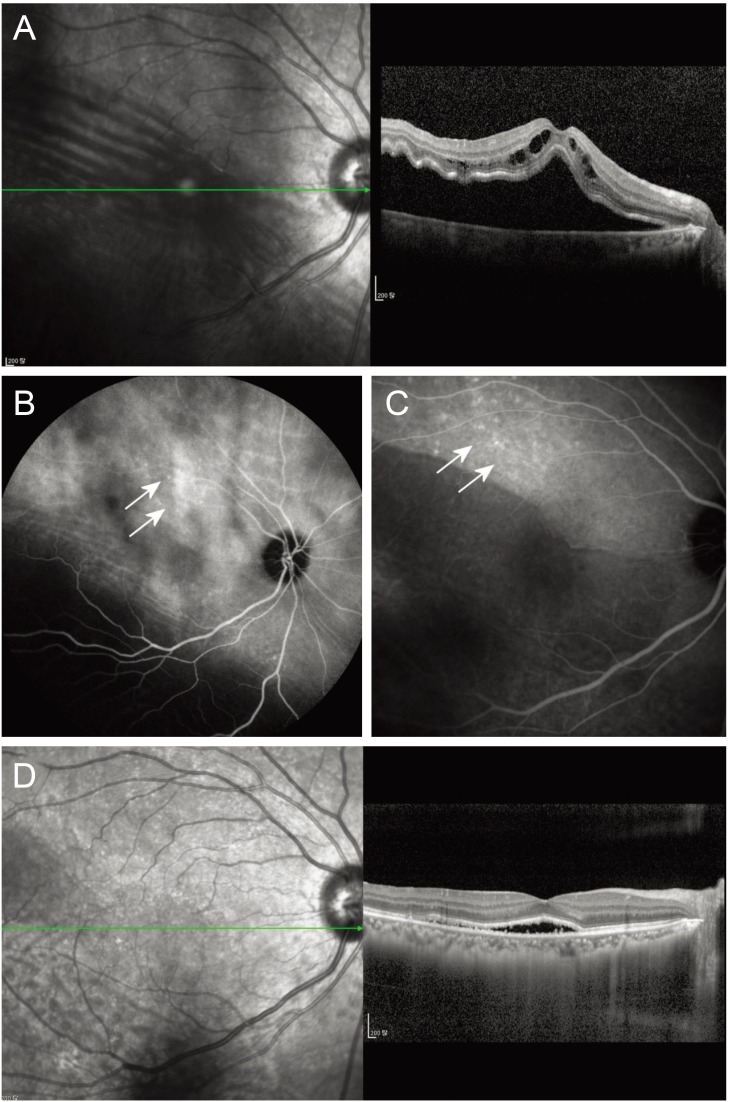
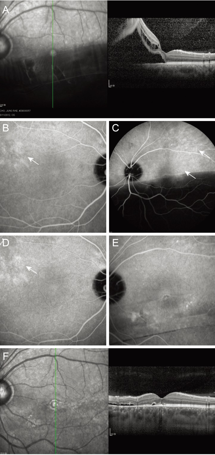
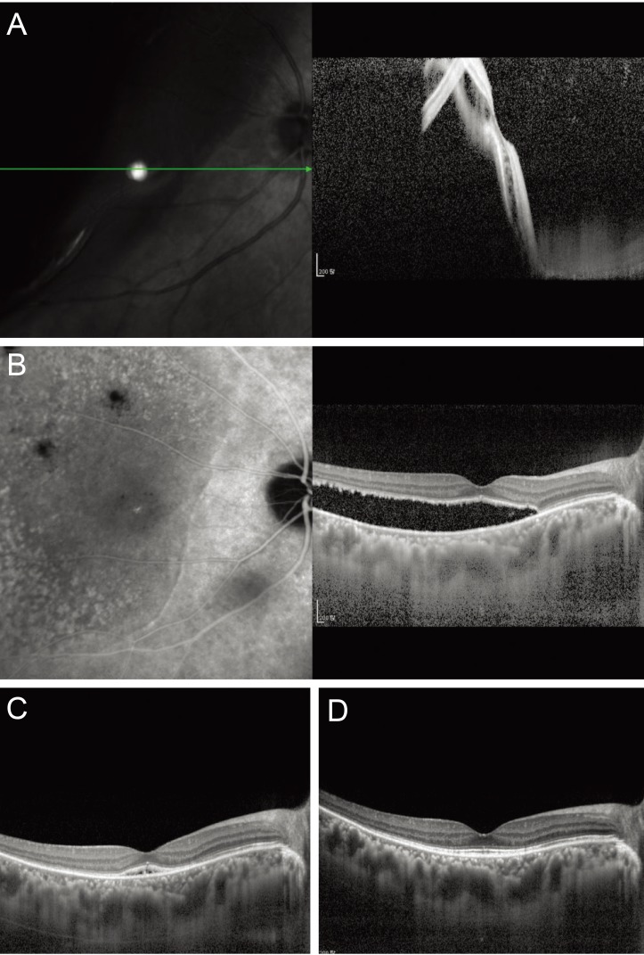
Methods: This study involved 36 eyes of 36 consecutive patients who underwent successful surgery for rhegmatogenous retinal detachment. A complete ophthalmologic evaluation, including clinical fundus examination, optical coherence tomography, and indocyanine green angiography, was conducted before and after surgery. Delayed absorption was defined as the presence of residual concave SRF or an SRF bleb at 6 months after surgery. Clinical factors and choroidal features on indocyanine green angiography were compared according to the presence and absence of delayed absorption.
Results: Eighteen of 36 eyes (50%) showed delayed absorption. Macular involvement and worse preoperative visual acuity were significantly related to the presence of delayed absorption (p = 0.001 and p = 0.034, respectively). On indocyanine green angiography, preoperative choroidal vascular hyperpermeability was noted in 70% of eyes with delayed absorption and in 14% of eyes without it (p = 0.010).
Conclusions: Delayed absorption of SRF after retinal reattachment surgery was not rare, with a 50% of incidence in this study. Macula-off status was significantly related to the incidence of delayed SRF absorption, and choroidal features such as choroidal vascular hyperpermeability might be responsible in part, possibly through the resultant exudative property of choroid.
Keywords: Angiography; Choroidal vascular hyperpermeability; Optical coherence tomography; Rhegmatogenous retinal detachment.
© 2017 The Korean Ophthalmological Society
Conflict of interest statement
No potential conflict of interest relevant to this article was reported.
Figures
References
-
- Machemer R. Experimental retinal detachment in the owl monkey. IV. The reattached retina. Am J Ophthalmol. 1968;66:1075–1091. - PubMed
-
- Baba T, Hirose A, Moriyama M, Mochizuki M. Tomographic image and visual recovery of acute macula-off rhegmatogenous retinal detachment. Graefes Arch Clin Exp Ophthalmol. 2004;242:576–581. - PubMed
-
- Benson SE, Schlottmann PG, Bunce C, et al. Optical coherence tomography analysis of the macula after vitrectomy surgery for retinal detachment. Ophthalmology. 2006;113:1179–1183. - PubMed
-
- Hagimura N, Iida T, Suto K, Kishi S. Persistent foveal retinal detachment after successful rhegmatogenous retinal detachment surgery. Am J Ophthalmol. 2002;133:516–520. - PubMed
-
- Kaga T, Fonseca RA, Dantas MA, et al. Optical coherence tomography of bleb-like subretinal lesions after retinal reattachment surgery. Am J Ophthalmol. 2001;132:120–121. - PubMed
MeSH terms
LinkOut - more resources
Full Text Sources
Other Literature Sources
Medical
Miscellaneous
