Case Reports
. 2018 Apr;20(4):464-469.
doi: 10.1038/gim.2017.128. Epub 2017 Sep 14.

Looking beyond the exome: a phenotype-first approach to molecular diagnostic resolution in rare and undiagnosed diseases

Collaborators, Affiliations
Case Reports

Looking beyond the exome: a phenotype-first approach to molecular diagnostic resolution in rare and undiagnosed diseases

Loren D M Pena et al. Genet Med. 2018 Apr.

Abstract

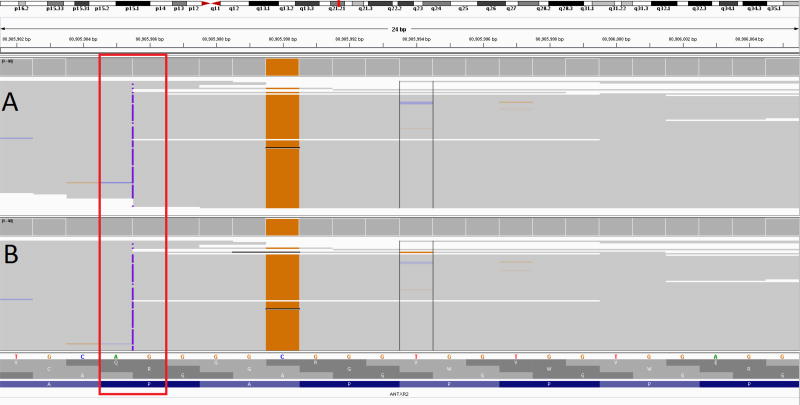
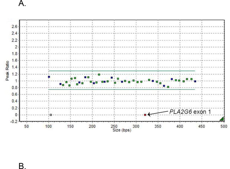
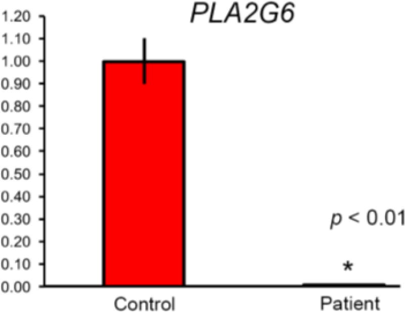
PurposeTo describe examples of missed pathogenic variants on whole-exome sequencing (WES) and the importance of deep phenotyping for further diagnostic testing.MethodsGuided by phenotypic information, three children with negative WES underwent targeted single-gene testing.ResultsIndividual 1 had a clinical diagnosis consistent with infantile systemic hyalinosis, although WES and a next-generation sequencing (NGS)-based ANTXR2 test were negative. Sanger sequencing of ANTXR2 revealed a homozygous single base pair insertion, previously missed by the WES variant caller software. Individual 2 had neurodevelopmental regression and cerebellar atrophy, with no diagnosis on WES. New clinical findings prompted Sanger sequencing and copy number testing of PLA2G6. A novel homozygous deletion of the noncoding exon 1 (not included in the WES capture kit) was detected, with extension into the promoter, confirming the clinical suspicion of infantile neuroaxonal dystrophy. Individual 3 had progressive ataxia, spasticity, and magnetic resonance image changes of vanishing white matter leukoencephalopathy. An NGS leukodystrophy gene panel and WES showed a heterozygous pathogenic variant in EIF2B5; no deletions/duplications were detected. Sanger sequencing of EIF2B5 showed a frameshift indel, probably missed owing to failure of alignment.ConclusionThese cases illustrate potential pitfalls of WES/NGS testing and the importance of phenotype-guided molecular testing in yielding diagnoses.


Conflict of interest statement

Conflicts of Interest

The rest of the authors declare no conflicts of interest related to this manuscript.

Figures

Figure 1
Figure 1
Integrated Genome Variant browser showing condensed read alignment for internally realigned data (A), and the BAM file provided by the clinical laboratory (B). Reads are shown stacked together, with colors indicating a mismatch to the reference sequence. Almost all reads show the pathogenic insertion (c.1073dupC, 22:g.80905986dupG) as a purple mark. The insertion call may be considered low quality because of the adjacent homopolymer repeat and nearby variant. The nearby SNP (c.1069G>C, 22:g.80905990C>G) can be seen by the majority of reads containing the orange G nucleotide change. This variant can also contribute to lower mapping quality and can affect variant quality at the insertion.
Figure 2
Figure 2
A. MLPA analysis of PLA2G6. Normalized MLPA data showing the homozygous deletion of the non-coding exon 1 of the PLA2G6 gene leading to total absence of amplification and hence a ratio of zero for the probe covering this region B. qRT-PCR analysis of PLA2G6 mRNA expression in blood. Results are expressed as means ± SD; mRNA levels were quantitated with real-time PCR and normalized to the level of GAPDH. The figure represents real-time PCR quantification of PLA2G6 gene expression in the patient compared to controls. All results were done in triplicates. P-value was calculated by student t-test.
Figure 2
Figure 2
A. MLPA analysis of PLA2G6. Normalized MLPA data showing the homozygous deletion of the non-coding exon 1 of the PLA2G6 gene leading to total absence of amplification and hence a ratio of zero for the probe covering this region B. qRT-PCR analysis of PLA2G6 mRNA expression in blood. Results are expressed as means ± SD; mRNA levels were quantitated with real-time PCR and normalized to the level of GAPDH. The figure represents real-time PCR quantification of PLA2G6 gene expression in the patient compared to controls. All results were done in triplicates. P-value was calculated by student t-test.

References

    1. Chong JX, Buckingham KJ, Jhangiani SN, et al. The Genetic Basis of Mendelian Phenotypes: Discoveries, Challenges, and Opportunities. Am J Hum Genet. 2015 - PMC - PubMed
    1. Need AC, Shashi V, Hitomi Y, et al. Clinical application of exome sequencing in undiagnosed genetic conditions. J Med Genet. 2012;49(6):353–361. - PMC - PubMed
    1. Yang Y, Muzny DM, Xia F, et al. Molecular findings among patients referred for clinical whole-exome sequencing. JAMA. 2014 - PMC - PubMed
    1. Lee H, Deignan JL, Dorrani N, et al. Clinical exome sequencing for genetic identification of rare mendelian disorders. JAMA. 2014 - PMC - PubMed
    1. El-Kamah GY, Fong K, El-Ruby M, et al. Spectrum of mutations in the ANTXR2 (CMG2) gene in infantile systemic hyalinosis and juvenile hyaline fibromatosis. Br J Dermatol. 2010;163(1):213–215. - PubMed

Publication types

MeSH terms