. 2018 Jan;37(1):117-125.
doi: 10.1007/s10067-017-3832-1. Epub 2017 Sep 15.

Thioredoxin 1 is associated with the proliferation and apoptosis of rheumatoid arthritis fibroblast-like synoviocytes

Affiliations

Thioredoxin 1 is associated with the proliferation and apoptosis of rheumatoid arthritis fibroblast-like synoviocytes

Tianbao Lu et al. Clin Rheumatol. 2018 Jan.

Abstract

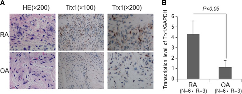
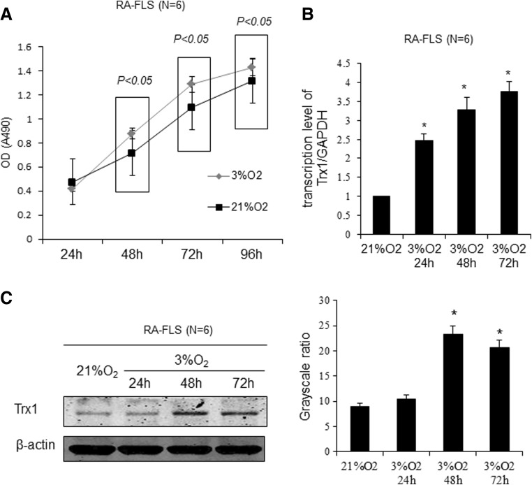
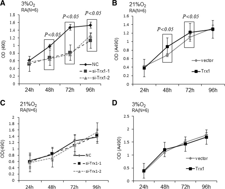
We aimed to investigate the possible effects of thioredoxin 1 (Trx1) on the proliferation and apoptosis of rheumatoid arthritis fibroblast-like synoviocytes (RA-FLSs) and elucidate the possible mechanisms involved. We investigated the distribution and expression of Trx1 in synovial tissues from RA and osteoarthritis (OA) patients by immunohistochemistry and real-time polymerase chain reaction (RT-PCR) analyses. RA-FLSs were isolated and cultured under normoxic (21% oxygen) or hypoxic (3% oxygen) concentrations. Transfection of Trx1-siRNAs and a Trx1 overexpression construct was conducted to manipulate the expression of Trx1. Protein expression was detected by Western blot. Doxorubicin (Adriamycin, ADR) was used to induce apoptosis. LY-294002 was used for the inhibition of PI3K-Akt. Cell proliferation and apoptosis were determined by MTS (3-[4,5-dimethylthiazol-2-yl]-5-[3-carboxymethoxyphenyl]-2-[4-sulfophenyl]-2H-tetrazolium, inner salt) assay and flow cytometry, respectively. The mRNA and protein expression of Trx1 in RA tissues was higher than that in OA tissues. The expression levels of Trx1 and cell proliferation in RA-FLSs were increased under hypoxia in comparison to those under normoxia. In hypoxia, downregulation of Trx1 significantly suppressed FLS proliferation, and the expression of PI3Kp85, phospho-Akt, and Bcl-2, while notably increased FLS apoptosis and the expression of active Caspase3 and Bax. In normoxia, Trx1 overexpression promoted the FLS proliferation and the expression of PI3Kp85, phospho-Akt, and Bcl-2, but inhibited FLS apoptosis and the expression of active Caspase3 and Bax in FLSs. Such effects were partially repressed by LY-294002 treatment. Trx1 may play an important role in regulating the proliferation and apoptosis of RA-FLSs by modulating PI3K-Akt activation.

Keywords: Apoptosis; Fibroblast-like synoviocytes; Proliferation; Rheumatoid arthritis; Thioredoxin 1.


Conflict of interest statement

None.

Figures

Fig. 1
Fig. 1
Trx1 is highly expressed in RA synovial tissues. a Immunohistochemical staining of Trx1 in synovium from RA and OA patients. b The mRNA levels of Trx1 in synovial tissues by quantitative RT-PCR. The data in b are means ± SD obtained from three separate experiments
Fig. 2
Fig. 2
Hypoxia promotes RA-FLS proliferation and induces Trx1 expression in FLSs. a Effect of different oxygen concentrations on cell proliferation as assessed by MTS assay. b Trx1 mRNA expression levels were measured by quantitative RT-PCR analysis in RA-FLSs. c Trx1 protein expression levels were evaluated by Western blot analysis in RA-FLSs. The data are showed as means ± SD. (*P < 0.05 compared with the group that was cultured under 21% O2)
Fig. 3
Fig. 3
Trx1 affects the proliferation of RA-FLSs. a, b RA-FLSs were transfected with Trx1-siRNAs (si-Trx1-1 and si-Trx1-2) or control siRNA (NC) and cultured under 3% O2 (a) and 21% O2 (b) condition. The growth curve of RA-FLSs under 3% O2 condition was determined by MTS assay. c, d RA-FLSs were transfected with a Trx1 expression construct or control vector and cultured under 21% O2 condition. The growth curve of RA-FLS under 21% O2 (c) and 3% O2 (d) condition was assessed by MTS assay. The data are shown as means ± SD
Fig. 4
Fig. 4
Trx1 affects the apoptosis of RA-FLSs. a RA-FLSs were transfected with Trx1-siRNAs (si-Trx1-1 and si-Trx1-2) or control siRNA (NC) and cultured for 48 h under 3% O2 and 21% O2 conditions. Cell apoptosis was then detected by flow cytometry. b Bar graph data of the early apoptotic cells (lower-right quadrant) are shown. c RA-FLSs were transfected with a Trx1 expression construct or control vector. After 48-h culture under 3% O2 and 21% O2 conditions, FLSs were incubated in the presence of 1 μg/mL of Doxorubicin (Adriamycin, ADR; Sangon, Shanghai, China) for 24 h. Cell apoptosis was then detected by flow cytometry. d Bar graph data of the early apoptotic cells (lower-right quadrant) are shown. The data in a and c are representatives of three separate experiments, and in b and d are shown as mean ± SD (*P < 0.05, **P < 0.01)
Fig. 5
Fig. 5
Trx1 modulates PI3K-Akt activation and the expression apoptosis-related proteins in RA-FLSs. a, c RA-FLSs were transfected with Trx1-siRNAs (si-Trx1-1 and si-Trx1-2) or control siRNA (NC) and cultured for 48 h under 3% O2 condition. Western blot and grayscale ratio analyses for the activation of PI3K-Akt and the expression of apoptosis-related proteins in RA-FLSs are shown. *P < 0.05. b, d RA-FLSs were transfected with a Trx1 expression construct or control vector. After 48-h culture under 21% O2 condition, 20 μM LY-294002 (Beyotime Biotechnology, Jiangsu, China) or an equal volume of DMSO was added to the cultured media. Cells were then incubated under normoxia for 24 h and Western blot analyses were then performed. Representative blot and grayscale ratio analyses of three separate experiments are shown (#P < 0.05 versus vector-transfected cells, ☆P < 0.05 Trx1-transfected cells versus Trx1-transfected and LY-294002-treated cell)

References

    1. Bottini N, Firestein GS. Duality of fibroblast-like synoviocytes in RA: passive responders and imprinted aggressors. Nat Rev Rheumatol. 2012;9(1):24–33. doi: 10.1038/nrrheum.2012.190. - DOI - PMC - PubMed
    1. Bartok B, Firestein GS. Fibroblast-like synoviocytes: key effector cells in rheumatoid arthritis. Immunol Rev. 2010;233(1):233–255. doi: 10.1111/j.0105-2896.2009.00859.x. - DOI - PMC - PubMed
    1. Ganesan R, Rasool M. Fibroblast-like synoviocytes-dependent effector molecules as a critical mediator for rheumatoid arthritis: current status and future directions. Int Rev Immunol. 2017;36(1):20–30. doi: 10.1080/08830185.2016.1269175. - DOI - PubMed
    1. Muz B, Khan MN, Kiriakidis S, Paleolog EM. Hypoxia. The role of hypoxia and HIF-dependent signalling events in rheumatoid arthritis. Arthritis Res Ther. 2009;11(1):201. doi: 10.1186/ar2568. - DOI - PMC - PubMed
    1. Juranek I, Stern R, Soltes L. Hyaluronan peroxidation is required for normal synovial function: an hypothesis. Med Hypotheses. 2014;82(6):662–666. doi: 10.1016/j.mehy.2014.02.024. - DOI - PubMed