Review
. 2017 Sep 15;22(9):1557.
doi: 10.3390/molecules22091557.

Probing Gas Adsorption in Zeolites by Variable-Temperature IR Spectroscopy: An Overview of Current Research

Affiliations
Review

Probing Gas Adsorption in Zeolites by Variable-Temperature IR Spectroscopy: An Overview of Current Research

Edoardo Garrone et al. Molecules. .

Abstract

The current state of the art in the application of variable-temperature IR (VTIR) spectroscopy to the study of (i) adsorption sites in zeolites, including dual cation sites; (ii) the structure of adsorption complexes and (iii) gas-solid interaction energy is reviewed. The main focus is placed on the potential use of zeolites for gas separation, purification and transport, but possible extension to the field of heterogeneous catalysis is also envisaged. A critical comparison with classical IR spectroscopy and adsorption calorimetry shows that the main merits of VTIR spectroscopy are (i) its ability to provide simultaneously the spectroscopic signature of the adsorption complex and the standard enthalpy change involved in the adsorption process; and (ii) the enhanced potential of VTIR to be site specific in favorable cases.

Keywords: IR spectroscopy; VTIR spectroscopy; dual sites; gas adsorption; zeolites.


Conflict of interest statement

The authors declare no conflict of interests.

Figures

Figure 1
Figure 1
Scheme of the homemade (stainless-steel) variable-temperature IR cell: (1) sample wafer, (2) sample holder, (3) magnetically-driven anchoring piece, (4) quartz tube, (5) hook for fixing the sample wafer inside the furnace, (6) furnace, (7) Viton O-ring, (8) cell body, (9) refrigerated region, (10) and (11) optical windows, (12) indium gaskets, (13) valve, (14) Teflon gasket, (15) pressure gauge ([33]).
Figure 2
Figure 2
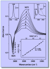
Representative variable-temperature IR spectra (O−H stretching region) of CO2 adsorbed on H-MCM-22. The spectra are shown in the difference mode (zeolite blank subtracted). From 1–7, temperature goes from 233–275 K; and equilibrium pressure from 4.49–6.22 mbar. The top inset (right) shows the IR spectra in the O−H stretching region of the blank zeolite H-MCM-22 wafer (black line), and after dosing with CO2 at 77 K (gray line). The top inset (left) shows the asymmetric ν3(CO2) stretching region of Spectrum 1. The bottom inset shows the van ’t Hoff plot for CO2 adsorbed on H-MCM-22: data obtained from the O−H stretching band at 3625 cm−1. R, linear regression coefficient; SD, standard deviation.
Figure 3
Figure 3
Adsorption heat of CO2 on H-MCM-22, at 303 K, as a function of coverage [35].
Figure 4
Figure 4
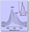
Representative variable-temperature IR spectra of CO adsorbed on Li-ZSM-5 (Si:Al = 30:1). From top to bottom, the temperature increases from 234–282 K; and equilibrium pressure from 3.85–5.01 mbar. The inset shows an example of the band resolution.
Figure 5
Figure 5
Van ’t Hoff plots for CO adsorbed on Li-ZSM-5 (Si:Al = 30:1); data obtained from the IR absorption bands at 2194 (LiA+···CO) and 2187 cm−1 (LiB+···CO). R, linear regression coefficient; SD, standard deviation.
Figure 6
Figure 6
Differential heat of adsorption of CO on Li-ZSM-5 at 303 K as a function of adsorbed amount. Black symbols: primary isotherm; empty symbols: secondary isotherm.
Figure 7
Figure 7
VTIR spectra (zeolite blank subtracted) of CO adsorbed on Na-FER, Si:Al = 8:1. Temperature increasing from 207–264 K; and equilibrium pressure from 0.19–1.17 mbar (from back to front).
Figure 8
Figure 8
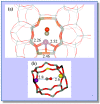
(a) Coordination of the Na+ cation and corresponding CO adsorption complex in a single cation site of Na-FER. Framework Al, Si and O atoms are depicted in black, grey and red colors, respectively; Na+, C and O atoms of CO are shown as violet, grey and red balls, respectively. The distances between Na+ and the nearest framework O atoms are given in Å; (b) CO adsorption complex on a dual cation site in Na-FER. The CO molecule interacts with the primary Na+ cation (violet ball) through the C atom (grey ball) and with the secondary Na+ cation (yellow ball) through the O atom (red ball). For further details, see [44].
Figure 9
Figure 9
Van ’t Hoff plots for CO adsorbed on Na-FER, data obtained from the IR absorption bands at 2158 cm−1 (Na+···CO···Na+) and at 2175 cm−1 (Na+···CO).
Figure 10
Figure 10
(a) T-shaped CO adsorption complex formed on the multiple cation site S2-(S1,S1) in Na-A. C and O atoms of the CO molecule are shown as grey and red spheres, respectively. Na+ cations in S1, S2 and S3 are depicted as white, blue and black balls, respectively; Na+ cations close to the CO molecule are shown as larger spheres; (b) Structure of a CO adsorption complex involving three cations in Na-CHA. Distances between primary and secondary cations and the CO molecule are given in Å. For details, see [59,60].
Figure 11
Figure 11
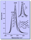
Representative variable-temperature IR (VTIR) spectra (ν3 region) of CO2 adsorbed on K-FER (Si:Al = 27.5:1). From 16, the temperature goes from 268–301 K; and equilibrium pressure from 0.21–0.50 mbar. The zeolite blank spectrum was subtracted. The top inset shows the IR spectra of increasing doses of CO2 adsorbed on K-FER (Si:Al = 8.6:1) at room temperature. The bottom inset shows van ’t Hoff plots obtained for IR absorption bands arising from the single site K+···OCO (band at 2346 cm−1) and the dual site K+···OCO···K+ (band at 2355 cm−1) adsorption complexes.

References

    1. Sumida K., Rogow D.L., Mason J.A., McDonald T.M., Bloch E.D., Herm Z.R., Bae T.H., Long J.R. Carbon dioxide capture in metal-organic frameworks. Chem. Rev. 2012;112:724–781. doi: 10.1021/cr2003272. - DOI - PubMed
    1. Wang Q.A., Luo J.Z., Zhong Z.Y., Borgna A. CO2 capture by solid adsorbents and their applications: Current status and new trends. Energy Environ. Sci. 2011;4:42–55. doi: 10.1039/C0EE00064G. - DOI
    1. Yazaydin A.O., Snurr R.Q., Park T.H., Koh K., Liu J., LeVan M.D., Benin A.I., Jakubeczak P., Lanuza M., Galloway D.B., et al. Screening metal-organic frameworks for carbon dioxide capture from flue gas using a combined experimental and modeling approach. J. Am. Chem. Soc. 2009;131:18198–18199. doi: 10.1021/ja9057234. - DOI - PubMed
    1. Dietzel P.D.C., Johnsen R.E., Fjellvaj H., Bordiga S., Groppo E., Chavan S., Blom R. Adsorption properties and structure of CO2 adsorbed on open coordination sites of metal-organic framework Ni2(dhtp) from gas adsorption, IR spectroscopy and X-ray diffraction. Chem. Commun. 2008;7:5125–5127. doi: 10.1039/b810574j. - DOI - PubMed
    1. Gomez-Alvarez P., Calero S. Highly selective zeolite topologies for flue gas separation. Chem. Eur. J. 2016;22:18705–18708. doi: 10.1002/chem.201604009. - DOI - PubMed

LinkOut - more resources