. 2017 Dec;24(12):2161-2172.
doi: 10.1038/cdd.2017.136. Epub 2017 Sep 15.

MicroRNA-200c impairs uterine receptivity formation by targeting FUT4 and α1,3-fucosylation

Affiliations

MicroRNA-200c impairs uterine receptivity formation by targeting FUT4 and α1,3-fucosylation

Qin Zheng et al. Cell Death Differ. 2017 Dec.

Abstract

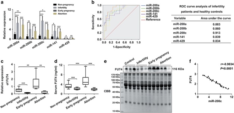
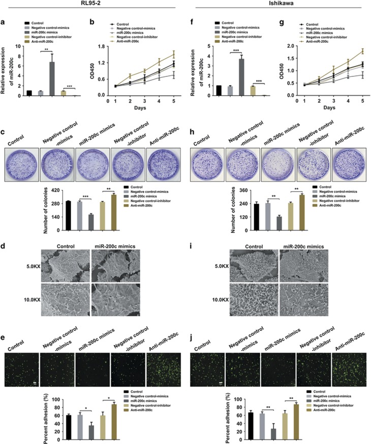
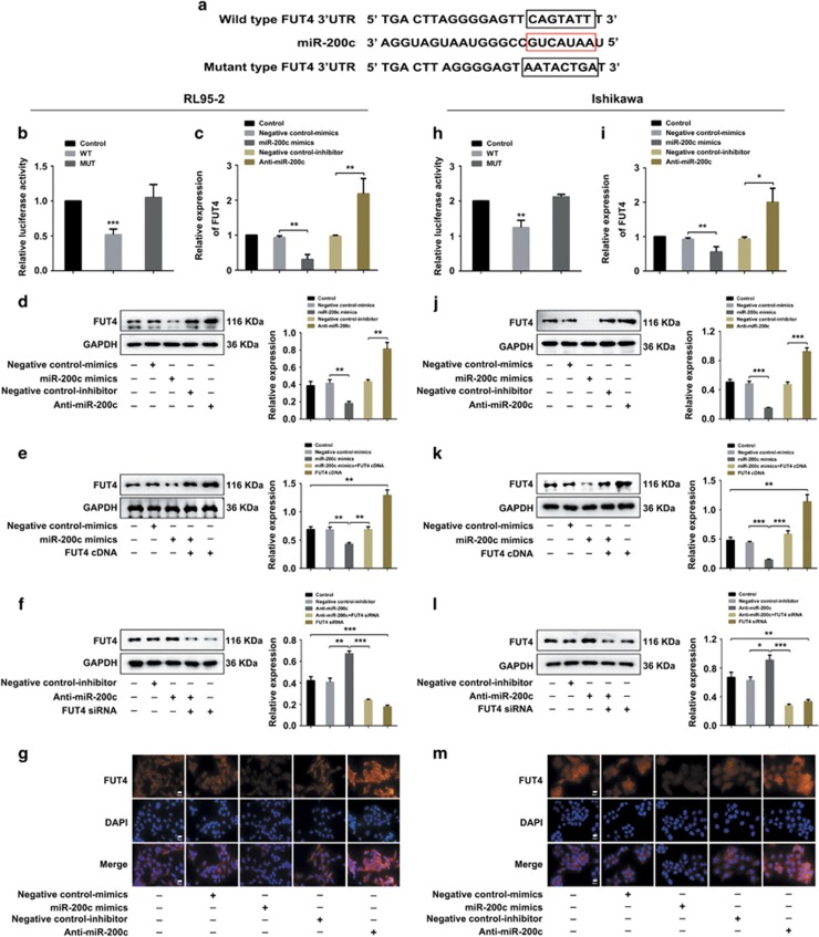
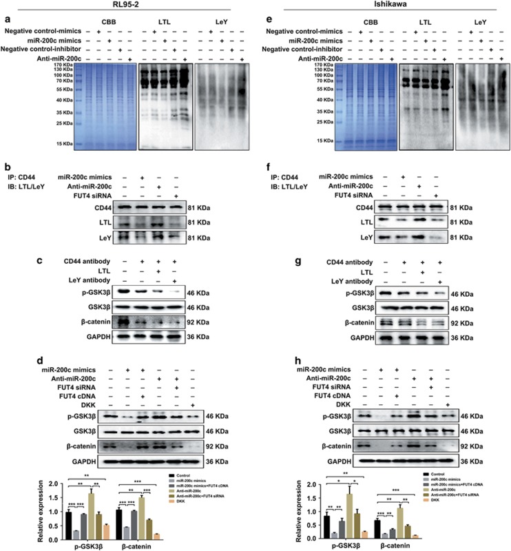
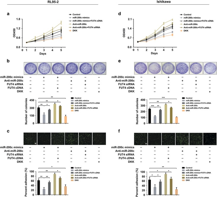
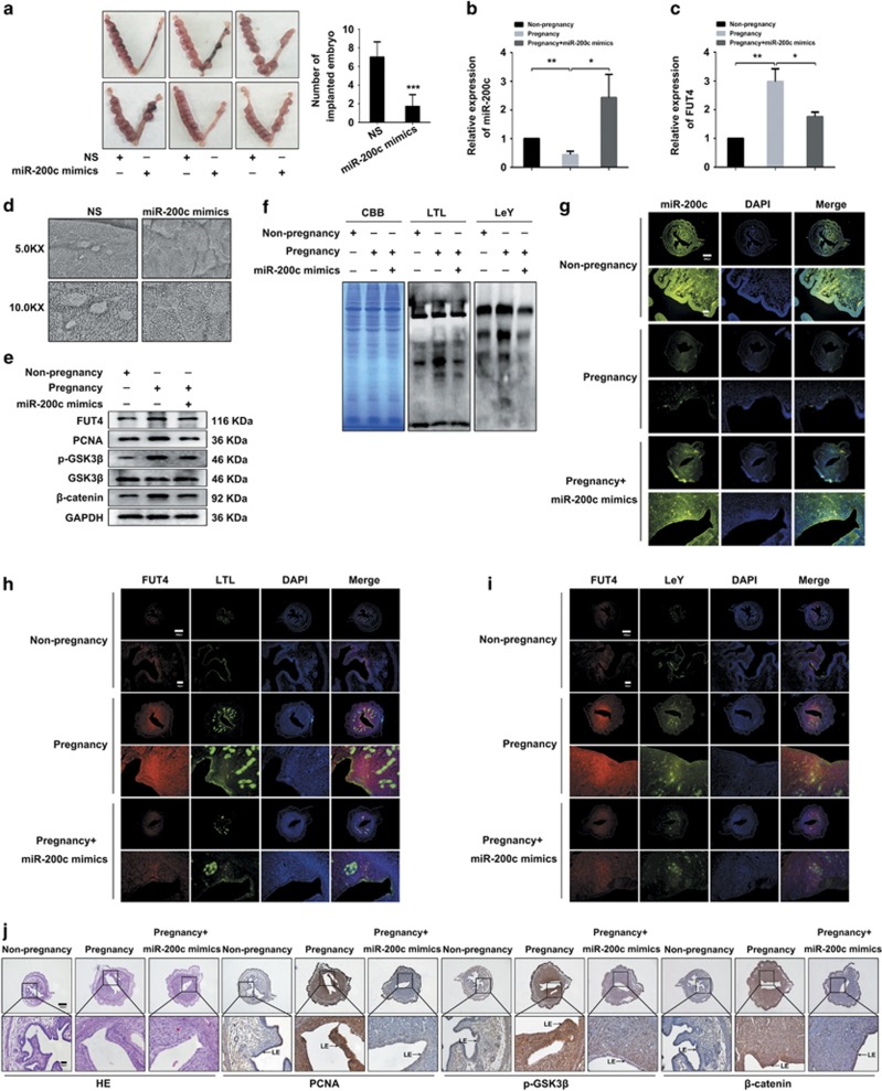
Successful embryo implantation requires the establishment of a receptive endometrium. Poor endometrial receptivity has generally been considered as a major cause of infertility. Protein glycosylation is associated with many physiological and pathological processes. The fucosylation is catalyzed by the specific fucosyltransferases. Fucosyltransferase IV (FUT4) is the key enzyme for the biosynthesis of α1,3-fucosylated glycans carried by glycoproteins, and the previous studies showed FUT4 expression changed dynamically during perimplantation. MicroRNAs (miRNAs) are known to regulate specific gene expression. However, the relationship between specific miRNA and FUT4, as well as the role of miRNA/FUT4 in the establishment of uterine receptivity remains elusive. In the current study, we reported that the levels of miR-200 family members were significantly increased in serum from infertility and abortion patients relative to healthy non-pregnancy and early-pregnancy women. Among these, miR-200c was the most sensitive diagnostic criterion for infertility by receiver operating characteristic curve analysis. FUT4 was lower in the serum from infertility and abortion patients compared with the healthy non-pregnancy and early-pregnancy women. Using endometrial cell lines and a mouse model, we demonstrated that miR-200c targeted and inhibited FUT4 expression, leading to the dysfunction of uterine receptivity. Our results also revealed that miR-200c decreased α1.3-fucosylation on glycoprotein CD44, which further inactivated Wnt/β-catenin signaling pathway. Taken together, miR-200c hampers uterine receptivity formation by targeting FUT4 and α1.3-fucosylation on CD44. miR-200c and FUT4 may be applied together as the potential markers for endometrial receptivity, and useful diagnostic and therapeutic targets for infertility.


Conflict of interest statement

The authors declare no conflict of interest.

Figures

Figure 1
Figure 1
High miR-200 family and low FUT4 levels in infertility and abortion patients. (a) Serum levels of miR-200 family members in healthy control (Non-pregnancy), infertility, early pregnancy and abortion detected by real-time PCR. (b) ROC curve analysis of the diagnostic value of miR-200 family members for infertility. (c–e) Detection of FUT4 expression by real-time PCR (c), ELISA (d) and western blot (e). (f) Correlation analysis between miR-200c and FUT4 expression in serum of healthy control and infertility patients. ROC: receiver operating characteristic. CBB: coomassie brilliant blue. The statistical analysis was shown. *P<0.05, **P<0.01, ***P<0.001
Figure 2
Figure 2
miR-200c inhibits proliferation and receptive ability of uterine epithelial cells in vitro. (a, f) Real-time PCR analysis for miR-200c expression in RL95-2 (a) and Ishikawa (f) cells after miR-200c mimics and Anti-miR-200c transfection. (b, c, g, h) CCK-8 (b, g) and colony formation assay (c, h) were used to evaluate cell proliferation capabilities of RL95-2 and Ishikawa cells. (d, i) Representative pictures of scanning electron microscopy (SEM) in RL95-2 (d) and Ishikawa (i) cells after miR-200c mimics transfection. (e, j) Adhesion percent of embryonic cells (JAR) to RL95-2 (e) and Ishikawa (j) cells. The statistical analysis was shown. *P<0.05, **P<0.01, ***P<0.001
Figure 3
Figure 3
FUT4 is a novel target of miR-200c. (a) Schematic representing reporter constructs of WT-FUT4 3’-UTR (upper panel), MUT-FUT4 3’-UTR with a mutated miR-200c binding site (lower panel) and miR-200c sequence (middle panel). (b, h) target of miR-200c was identified by dual-luciferase gene reporter assay in both RL95-2 (b) and Ishikawa (h) cells. WT: wild-type FUT4 3’-UTR transfection; MUT: mutant-type FUT4 3’-UTR transfection. (c, d, i, j) Real-time PCR (c, i) and western blot analysis (d, j) of FUT4 level after miR-200c mimics and Anti-miR-200c transfection in uterine epithelial cells. The statistical analysis was shown. (e, k) Western blot analysis of FUT4 level in RL95-2 (e) and Ishikawa (k) cells transfected with miR-200c mimics or co-transfected with miR-200c mimics and FUT4 cDNA, respectively. The statistical analysis was shown. (f, l) Western blot analysis of FUT4 level in RL95-2 (f) and Ishikawa (l) cells transfected with Anti-miR-200c or co-transfected with Anti-miR-200c and FUT4 siRNA, respectively. The statistical analysis was shown. (g, m) Immunofluorescent staining of FUT4 after miR-200c mimics and Anti-miR-200c transfection in uterine epithelial cells. Scale bars, 20 μm. *P<0.05, **P<0.01, ***P<0.001
Figure 4
Figure 4
miR-200c decreases α1,3-fucosylation on CD44 and inactivates Wnt/β-catenin signaling pathway. (a, e) Western/lectin blot analysis of effect of miR-200c on α1,3-fucosylation and LeY biosynthesis in RL95-2 (a) and Ishikawa (e) cells. CBB: coomassie brilliant blue. LTL: Lotus tetragonolobus lectin. (b, f) Immunoprecipitation and western blot analysis of α 1,3-fucosylation and LeY on CD44 in RL95-2 (b) and Ishikawa (f) cells. Immunoprecipitation (IP): anti-CD44 antibody pulled down protein. Immune blot (IB): detection of α 1,3-fucosylation by LTL lectin and anti-LeY antibody. (c, g) Western blot analysis of CD44, LTL and LeY blocking on activation of p-GSK3β, GSK3β and β-catenin in RL95-2 (c) and Ishikawa (g) cells. (d, h) Western blot and statistical analysis of p-GSK3β, GSK3β and β-catenin in RL95-2 (d) and Ishikawa (h) cells. DKK: inhibitor of Wnt/β-catenin signal pathway. *P<0.05, **P<0.01, ***P<0.001
Figure 5
Figure 5
miR-200c inhibits proliferation and receptivity of uterine epithelial cells by targeting FUT4. (a, b, d, e) CCK-8 (a, d) and colony formation assay (b, e) were used to evaluate cell proliferation abilities of RL95-2 and Ishikawa cells. The statistical analysis was shown. (c, f) Adhesion percent of embryonic cells (JAR) to RL95-2 (c) and Ishikawa (f) cells. The statistical analysis was shown. *P<0.05, **P<0.01, ***P<0.001
Figure 6
Figure 6
miR-200c inhibits uterine receptivity formation and embryo implantation potential in vivo. Mouse uterus was injected with normal saline (NS) or miR-200c mimics on day 3 of pregnancy. (a) Number of implanted embryos in the uterus on day 9 pregnancy. The statistical analysis was shown. (b, c) Real-time PCR analysis of miR-200c (b) and FUT4 (c) in the uterine endometrium on day 4 of pregnancy. (d) Representative SEM images in the uterine endometrium after NS or miR-200c mimic injection on day 4 of pregnancy. (g) Fluorescent in situ hybridization (FISH) of miR-200c in the uterine endometrium on day 4 of pregnancy. (e, j) Western blot (e) and immunohistochemistry (j) analysis of PCNA and Wnt/β-catenin signaling pathway in the uterine endometrium of pregnancy mouse. (f, h, i) Lectin blot (f) and immunofluorescenct staining (h, i) detection of α1,3-fucosylation (LTL), LeY and FUT4 in the uterine endometrium of pregnancy mouse. LE: luminal epithelium. The statistical analysis was shown. *P<0.05, **P<0.01, ***P<0.001

Similar articles

Cited by

References

    1. Su RW, Fazleabas AT. Implantation and establishment of pregnancy in human and nonhuman primates. Adv Anat Embryol Cell Biol 2015; 216: 189–213. - PMC - PubMed
    1. Sharma A, Kumar P. Understanding implantation window, a crucial phenomenon. J Hum Reprod Sci 2012; 5: 2–6. - PMC - PubMed
    1. Bartosch C, Lopes JM, Beires J, Sousa M. Human endometrium ultrastructure during the implantation window: a new perspective of the epithelium cell types. Reprod Sci 2011; 18: 525–539. - PubMed
    1. Salamonsen LA, Evans J, Nguyen HP, Edgell TA. The microenvironment of human Implantation: determinant of reproductive success. Am J Reprod Immunol 2016; 75: 218–225. - PubMed
    1. Riad ON, Hak AA. Assessment of endometrial receptivity using Doppler ultrasonography in infertile women undergoing intrauterine insemination. Gynecol Endocrinol 2014; 30: 70–73. - PubMed

MeSH terms

LinkOut - more resources