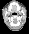
The accessory parotid gland and facial process of the parotid gland on computed tomography
- PMID: 28915265
- PMCID: PMC5600370
- DOI: 10.1371/journal.pone.0184633
The accessory parotid gland and facial process of the parotid gland on computed tomography
Abstract
The purpose of this study was to determine the incidence of an anterior extension of the parotid gland, such as an accessory parotid gland (APG) or facial process (FP) and to evaluate its characteristics on computed tomography (CT) scans. We reviewed CT scans of 1,600 parotid glands from 800 patients. An APG on CT was defined as a soft-tissue mass of the same density as the main parotid gland, located at the anterior part of the main parotid gland, and completely separate from the main parotid gland. An FP was defined as a lobe of the parotid gland protruding anteriorly over the anterior edge of the ramus of the mandible on CT and showing continuity with the main gland. The overall incidence rates and characteristics of APGs and FPs were evaluated according to age, sex, and side. The incidence rates of APGs and FPs were 10.2% (163/1,600) and 28.3% (452/1,600), respectively. The mean size of an APG was 15.8 mm × 5.0 mm and the mean distance from the main parotid gland was 10.5 mm. The FP reached anteriorly between the anterior edge of the mandibular ramus and the anterior border of the masseter muscle in 405 (89.6%) cases, while it extended over the anterior border of the masseter muscle in 47 (10.4%) cases. The incidence rates of APGs and FPs decreased and increased, respectively, with increasing age, showing significant linear correlations. However, the incidence of an anterior extension of the parotid gland (either an APG or an FP) was similar across all age groups. The present study showed that CT might be helpful in identifying anterior extensions of the parotid gland including APGs and FPs. The anatomical information gained from this study contributes to a better understanding of APGs and FPs and how their incidence changes with age.
Conflict of interest statement
Figures


References
-
- Frommer J. The human accessory parotid gland: its incidence, nature, and significance. Oral Surg Oral Med Oral Pathol. 1977;43(5):671–6. Epub 1977/05/01. . - PubMed
-
- Lin DT, Coppit GL, Burkey BB, Netterville JL. Tumors of the accessory lobe of the parotid gland: a 10-year experience. Laryngoscope. 2004;114(9):1652–5. Epub 2004/10/12. doi: 10.1097/00005537-200409000-00028 . - DOI - PubMed
-
- Newberry TR, Kaufmann CR, Miller FR. Review of accessory parotid gland tumors: pathologic incidence and surgical management. Am J Otolaryngol. 2014;35(1):48–52. Epub 2013/09/21. doi: 10.1016/j.amjoto.2013.08.018 . - DOI - PubMed
-
- Toh H, Kodama J, Fukuda J, Rittman B, Mackenzie I. Incidence and histology of human accessory parotid glands. Anat Rec. 1993;236(3):586–90. Epub 1993/07/01. doi: 10.1002/ar.1092360319 . - DOI - PubMed
-
- Klotz DA, Coniglio JU. Prudent management of the mid-cheek mass: revisiting the accessory parotid gland tumor. Laryngoscope. 2000;110(10 Pt 1):1627–32. Epub 2000/10/19. doi: 10.1097/00005537-200010000-00010 . - DOI - PubMed
MeSH terms
LinkOut - more resources
Full Text Sources
Other Literature Sources
Miscellaneous

