. 2017 Aug 3;8(34):55848-55862.
doi: 10.18632/oncotarget.19862. eCollection 2017 Aug 22.

Loss of p16INK4A stimulates aberrant mitochondrial biogenesis through a CDK4/Rb-independent pathway

Affiliations

Loss of p16INK4A stimulates aberrant mitochondrial biogenesis through a CDK4/Rb-independent pathway

Ethika Tyagi et al. Oncotarget. .

Abstract

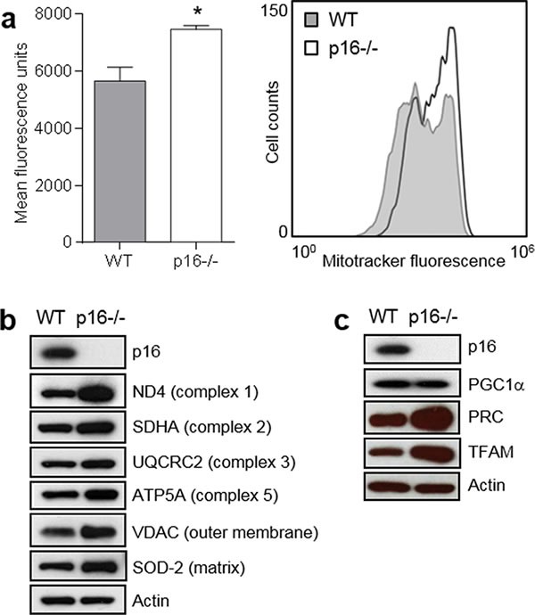
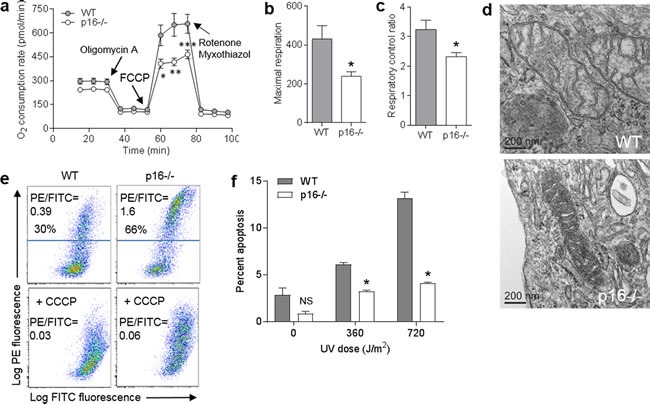
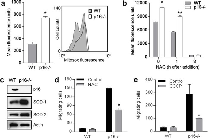
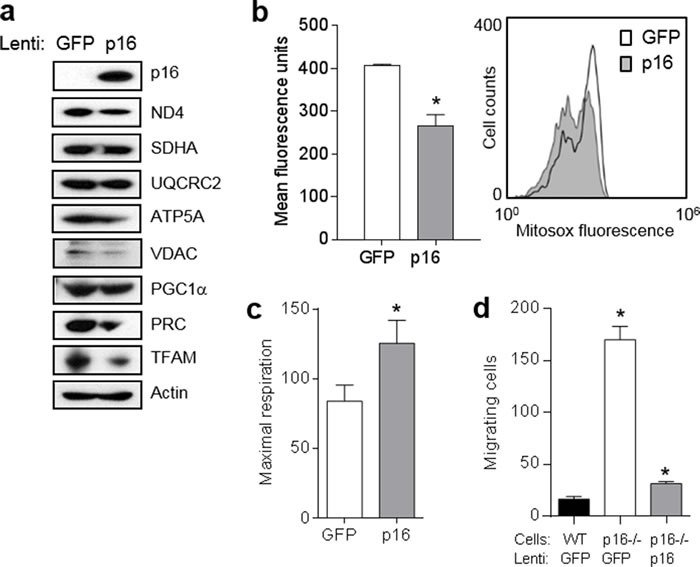
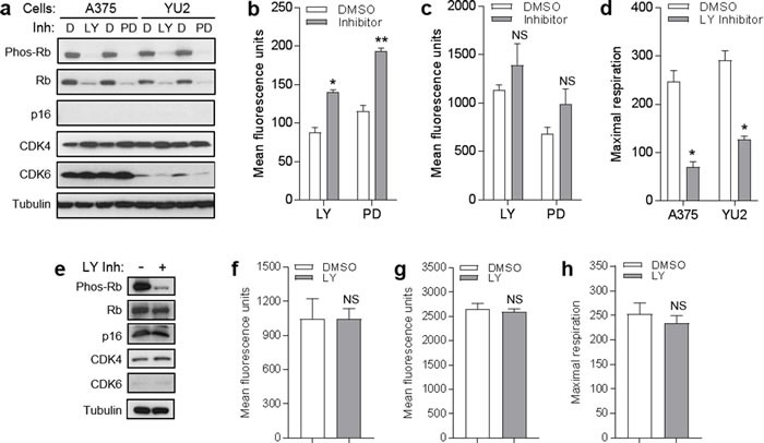
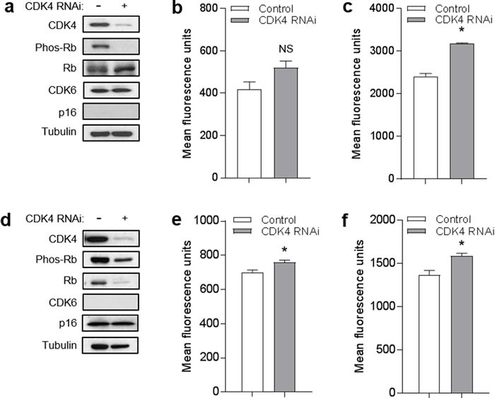
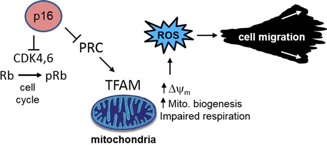
The tumor suppressor p16INK4A (p16) inhibits cell cycle progression through the CDK4/Rb pathway. We have previously shown that p16 regulates cellular oxidative stress, independent of its role in cell cycle control. We investigated whether loss of p16 had a direct impact on the mitochondria. We found that p16-null primary mouse fibroblasts (PMFs) displayed increased mitochondrial mass and expression of mitochondrial respiratory subunit proteins compared to wild-type (WT) PMFs. These findings in p16-null PMFs were associated with increased expression of the mitochondrial biogenesis transcription factors PRC and TFAM. On the other hand, p16-deficient PMFs demonstrated reduced mitochondrial respiration capacity consistent with electron microscopy findings showing that mitochondria in p16-deficient PMFs have abnormal morphology. Consistent with increased mitochondrial mass and reduced respiratory capacity, p16-deficient PMFs generated increased mitochondrial superoxide. One biological consequence of elevated ROS in p16-deficient PMFs was enhanced migration, which was reduced by the ROS scavenger N-acetylcysteine. Finally, p16-deficient PMFs displayed increased mitochondrial membrane potential, which was also required for their enhanced migration. The mitochondrial and migration phenotype was restored in p16-deficient PMFs by forced expression of p16. Similarly, over-expression of p16 in human melanocytes and A375 melanoma cells led to decreased expression of some mitochondrial respiratory proteins, enhanced respiration, and decreased migration. Inhibition of Rb phosphorylation in melanocytes and melanoma cells, either by addition of chemical CDK4 inhibitors or RNAi-mediated knockdown of CDK4, did not mimic the effects of p16 loss. These results suggest that p16 regulates mitochondrial biogenesis and function, which is independent of the canonical CDK4/Rb pathway.

Keywords: CDK4; fibroblast; migration; mitochondria; p16.


Conflict of interest statement

CONFLICTS OF INTEREST The authors declare that no conflicts of interest exist.

Figures

Figure 1
Figure 1. p16-deficient cells display increased mitochondrial mass and biogenesis
a. Mitochondrial mass. Left, wild-type (WT) and p16−/− primary fibroblasts were stained with MitoTracker-Green, and analyzed by flow cytometry. Error bars indicate SEM from triplicate determinations, *P = 0.02. Right, representative histogram. b. Western blotting for mitochondrial structural proteins in WT and p16−/− fibroblasts, with Actin as a loading control. c. Western blotting for proteins in WT and p16−/− fibroblasts that regulate mitochondrial biogenesis, with Actin as a loading control.
Figure 2
Figure 2. Loss of p16 results in decreased respiration capacity and mitochondrial changes
a. Oxygen consumption rate measured in wild-type (WT) and p16−/− primary fibroblasts with timed addition of oligomycin A (1 μg/ml), FCCP (0.5 μM), and a mixture of rotenone (1 μM) and myxothiazol (1 μM). Error bars indicate SEM from 9 determinations. *P = .02, **P = .007, ***P = .01. b. Maximal respiration. Values calculated from data in a.. *P = .02. c. Respiratory control ratio. Values calculated from data in a.. *P = .03. d. Representative electron micrographs of mitochondria. Scale bar indicates 200 nm. e. WT and p16−/− fibroblasts were stained with JC-1, in the absence or presence of 50 μM CCCP. f. WT and p16−/− fibroblasts were treated with the indicated doses of UV, then assessed for apoptosis by Annexin V staining 24 h later. Error bars indicate SEM from 3 determinations. *P < .001. NS, not significant.
Figure 3
Figure 3. Elevated mitochondrial superoxide in p16-deficient cells is associated with enhanced cell migration
a. Wild-type (WT) and p16−/− primary fibroblasts were stained with MitoSox-Red and assessed for mitochondrial superoxide by flow cytometry. Error bars indicate SEM from triplicate determinations, *P < .001. Representative histogram at right. b. Detection of superoxide in the absence (0 h) or presence of 5 mM N-acetylcysteine (NAC) after 1 or 24 h. Error bars indicate SEM from triplicate determinations, *P < .001, *P = .006. c. Western blots of lysates from cells in a.. d. Wild-type (WT) and p16−/− fibroblasts were subjected to transwell migration assay, in the absence or presence of 5 mM NAC, and migrating cells were counted at 24 h. Error bars indicate SEM from triplicate determinations, *P = .003. e. Migration assay as in d., with cells in absence or presence of 50 μM CCCP. Error bars indicate SEM from triplicate determinations, *P = .005.
Figure 4
Figure 4. Expression of p16 in p16-deficient fibroblasts restores mitochondrial dynamics and motility
a. Fibroblasts isolated from p16−/− mice were infected with control (GFP) or p16-expressing lentivirus, then 6 d later lysates were subjected to Western blotting. Representative of two experiments performed. b. Cells in a. after 6d were analyzed for mitochondrial superoxide by flow cytometry. Error bars indicate SEM from triplicate determinations, *P = .005. Representative histogram at right. c. Maximal respiration of cells in a.. Error bars indicate SEM from 9 determinations, *P = .05. d. Fibroblasts isolated from wild-type (WT) or p16−/− mice were infected with control (GFP) or p16-expressing lentivirus, then 48 h later were placed in migration assay, and migrating cells counted at 24 h. Error bars indicate SEM from triplicate determinations, *P < .001.
Figure 5
Figure 5. Forced expression of p16 modulates mitochondrial biogenesis, respiration, and motility
a. A375 human melanoma cells were infected with control (GFP) or GFP/p16-expressing lentivirus, then 6 d later lysates were subjected to Western blotting. b. Cells in a. analyzed for mitochondrial superoxide by flow cytometry. Error bars indicate SEM from duplicate determinations, *P < .001. c. Maximal respiration of cells in a.. Error bars indicate SEM from 9 determinations, *P = .04. d. A375 cells were infected with control (GFP) or GFP/p16-expressing lentivirus, then 48 h later were placed in migration assay, and migrating cells counted at 24 h. Error bars indicate SEM from triplicate determinations, *P < .001. e. Human melanocytes were infected with control (GFP) or GFP/p16-expressing lentivirus, then 6 d later lysates were subjected to Western blotting. f. Cells in e. analyzed for mitochondrial superoxide by flow cytometry. Error bars indicate SEM from duplicate determinations, *P < .001. g. Maximal respiration of cells in e.. Error bars indicate SEM from 9 determinations, *P < .001. h. Melanocytes were infected with control (GFP) or GFP/p16-expressing lentivirus, then 48 h later were placed in migration assay and migrating cells counted at 48 h. Error bars indicate SEM from triplicate determinations, *P = .003.
Figure 6
Figure 6. CDK4/6 inhibitors do not mimic effects of p16 on mitochondrial dynamics and respiration
a. A375 and YU2 melanoma cells were treated with DMSO control d., 1 μM LY-2835219 (LY), or 2 μM PD-0332991 (PD), then lysates were analyzed by Western blotting 24 h later. b. A375 cells in a. analyzed for mitochondrial superoxide by flow cytometry. Error bars indicate SEM from triplicate determinations; *P = .05, **P < .001. c. A375 cells in a. analyzed for mitochondrial mass by flow cytometry. Error bars indicate SEM from triplicate determinations; *P = .01. d. Maximal respiration of A375 and YU2 cells in a.. Error bars indicate SEM from 5 determinations, *P = . < .001. e. Human melanocytes were treated with 1 μM LY or DMSO control, then lysates were analyzed by Western blotting 24 h later. f. Cells in e. analyzed for mitochondrial superoxide by flow cytometry. Error bars indicate SEM from triplicate determinations; NS, not significant. g. Cells in e. analyzed for mitochondrial mass by flow cytometry. Error bars indicate SEM from triplicate determinations; NS, not significant. h. Maximal respiration of cells in e.. Error bars indicate SEM from 5 determinations; NS, not significant.
Figure 7
Figure 7. CDK4 knockdown does not mimic effects of p16 on mitochondrial dynamics
a. YU2 melanoma cells were transfected with control or RNAi specific for CDK4, then lysates were analyzed by Western blotting 72 h later. b. Cells in a. analyzed for mitochondrial superoxide by flow cytometry. Error bars indicate SEM from triplicate determinations; NS, not significant. c. Cells in a. analyzed for mitochondrial mass by flow cytometry. Error bars indicate SEM from triplicate determinations; *P < .001. d. Human melanocytes were transfected with control or RNAi specific for CDK4, then lysates were analyzed by Western blotting 72 h later. e. Cells in d. analyzed for mitochondrial superoxide by flow cytometry. Error bars indicate SEM from triplicate determinations; *P = .04. f. Cells in d. analyzed for mitochondrial mass by flow cytometry. Error bars indicate SEM from triplicate determinations; *P = .04.
Figure 8
Figure 8. p16 control of mitochondrial biogenesis, ROS, and cell migration
Loss of p16 results in aberrant mitochondrial biogenesis, characterized by increased mitochondrial mass but dysfunctional respiration which results in elevated ∆ψm and ROS. These functions are independent of the CDK4/Rb pathway and cell cycle control.

Similar articles

Cited by

References

    1. Okamoto A, Demetrick DJ, Spillare EA, Hagiwara K, Hussain SP, Bennett WP, Forrester K, Gerwin B, Serrano M, Beach DH, Harris CC. Mutations and altered expression of p16INK4 in human cancer. Proc Natl Acad Sci U S A. 1994;91:11045–11049. - PMC - PubMed
    1. Sharpless NE, DePinho RA. The INK4A/ARF locus and its two gene products. Curr Opin Genet Dev. 1999;9:22–30. - PubMed
    1. Rayess H, Wang MB, Srivatsan ES. Cellular senescence and tumor suppressor gene p16. Int J Cancer. 2012;130:1715–1725. - PMC - PubMed
    1. Zuo L, Weger J, Yang Q, Goldstein AM, Tucker MA, Walker GJ, Hayward N, Dracopoli NC. Germline mutations in the p16INK4a binding domain of CDK4 in familial melanoma. Nat Genet. 1996;12:97–99. - PubMed
    1. Hashemi J, Platz A, Ueno T, Stierner U, Ringborg U, Hansson J. CDKN2A germ-line mutations in individuals with multiple cutaneous melanomas. Cancer Res. 2000;60:6864–6867. - PubMed