The roles of ING5 in gliomas: a good marker for tumorigenesis and a potential target for gene therapy
- PMID: 28915612
- PMCID: PMC5593583
- DOI: 10.18632/oncotarget.17802
The roles of ING5 in gliomas: a good marker for tumorigenesis and a potential target for gene therapy
Abstract
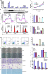
To elucidate the anti-tumor effects and molecular mechanisms of ING5 on glioma cells, we overexpressed it in U87 cells, and examined the phenotypes and their relevant molecules. It was found that ING5 overexpression suppressed proliferation, energy metabolism, migration, invasion, and induced G2/M arrest, apoptosis, dedifferentiation, senescence, mesenchymal- epithelial transition and chemoresistance to cisplatin, MG132, paclitaxel and SAHA in U87 cells. There appeared a lower expression of N-cadherin, Twist, Slug, Zeb1, Zeb2, Snail, Ac-H3, Ac-H4, Cdc2, Cdk4 and XIAP, but a higher expression of Claudin 1, Histones 3 and 4, p21, p53, Bax, β-catenin, PI3K, Akt, and p-Akt in ING5 transfectants. ING5 overexpression suppressed tumor growth of U87 cells in nude mice by inhibiting proliferation and inducing apoptosis. Down-regulated ING5 expression was closely linked to the tumorigenesis and histogenesis of glioma. These data indicated that ING5 expression might be considered as a good marker for the tumorigenesis and histogenesis of gliomas. It might be employed as a potential target for gene therapy of glioma. PI3K/Akt or β-catenin/TCF-4 activation might be positively linked to chemotherapeutic resistance, mediated by ING5.
Keywords: ING5; chemotherapy; gene therapy; glioma; tumorigenesis.
Conflict of interest statement
CONFLICTS OF INTEREST The authors declare that they have no competing interests.
Figures


Similar articles
-
The roles of ING5 in cancer: A tumor suppressor.Front Cell Dev Biol. 2022 Nov 8;10:1012179. doi: 10.3389/fcell.2022.1012179. eCollection 2022. Front Cell Dev Biol. 2022. PMID: 36425530 Free PMC article. Review.
-
ING5 suppresses proliferation, apoptosis, migration and invasion, and induces autophagy and differentiation of gastric cancer cells: a good marker for carcinogenesis and subsequent progression.Oncotarget. 2015 Aug 14;6(23):19552-79. doi: 10.18632/oncotarget.3735. Oncotarget. 2015. PMID: 25980581 Free PMC article.
-
The down-regulated ING5 expression in lung cancer: a potential target of gene therapy.Oncotarget. 2016 Aug 23;7(34):54596-54615. doi: 10.18632/oncotarget.10519. Oncotarget. 2016. PMID: 27409347 Free PMC article.
-
The nucleocytoplasmic translocation and up-regulation of ING5 protein in breast cancer: a potential target for gene therapy.Oncotarget. 2017 May 17;8(47):81953-81966. doi: 10.18632/oncotarget.17918. eCollection 2017 Oct 10. Oncotarget. 2017. PMID: 29137236 Free PMC article.
-
The roles of ING5 expression in ovarian carcinogenesis and subsequent progression: a target of gene therapy.Oncotarget. 2017 Oct 19;8(61):103449-103464. doi: 10.18632/oncotarget.21968. eCollection 2017 Nov 28. Oncotarget. 2017. PMID: 29262575 Free PMC article.
Cited by
-
Involvement of Bcl-2 Family Proteins in Tetraploidization-Related Senescence.Int J Mol Sci. 2023 Mar 28;24(7):6374. doi: 10.3390/ijms24076374. Int J Mol Sci. 2023. PMID: 37047342 Free PMC article. Review.
-
Inhibitor of Growth Factors Regulate Cellular Senescence.Cancers (Basel). 2022 Jun 24;14(13):3107. doi: 10.3390/cancers14133107. Cancers (Basel). 2022. PMID: 35804879 Free PMC article. Review.
-
ING5 activity in self-renewal of glioblastoma stem cells via calcium and follicle stimulating hormone pathways.Oncogene. 2018 Jan 18;37(3):286-301. doi: 10.1038/onc.2017.324. Epub 2017 Sep 18. Oncogene. 2018. PMID: 28925404 Free PMC article.
-
The Antitumor and Sorafenib-resistant Reversal Effects of Ursolic Acid on Hepatocellular Carcinoma via Targeting ING5.Int J Biol Sci. 2024 Aug 5;20(11):4190-4208. doi: 10.7150/ijbs.97720. eCollection 2024. Int J Biol Sci. 2024. PMID: 39247819 Free PMC article.
-
The roles of ING5 in cancer: A tumor suppressor.Front Cell Dev Biol. 2022 Nov 8;10:1012179. doi: 10.3389/fcell.2022.1012179. eCollection 2022. Front Cell Dev Biol. 2022. PMID: 36425530 Free PMC article. Review.
References
-
- Soliman MA, Riabowol K. After a decade of study-ING, a PHD for a versatile family of proteins. Trends Biochem Sci. 2007;32:509–519. - PubMed
LinkOut - more resources
Full Text Sources
Other Literature Sources
Research Materials
Miscellaneous

