. 2017 May 26;8(34):56816-56828.
doi: 10.18632/oncotarget.18232. eCollection 2017 Aug 22.

Selection of optimal molecular targets for tumor-specific imaging in pancreatic ductal adenocarcinoma

Affiliations

Selection of optimal molecular targets for tumor-specific imaging in pancreatic ductal adenocarcinoma

Willemieke S Tummers et al. Oncotarget. .

Abstract

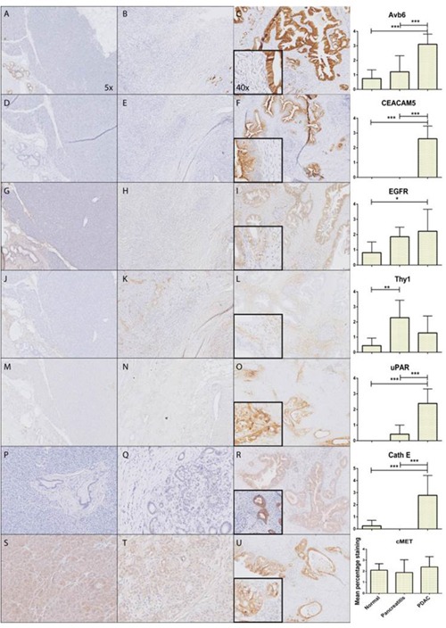
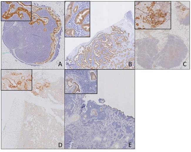
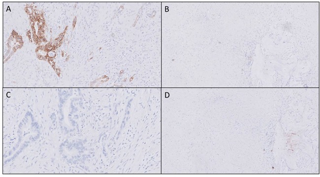
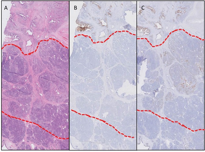
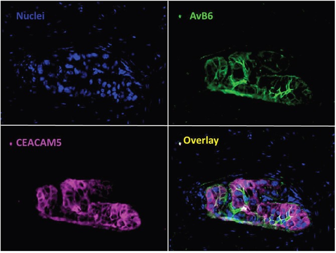
Discrimination of pancreatic ductal adenocarcinoma (PDAC) from chronic pancreatitis (CP) or peritumoral inflammation is challenging, both at preoperative imaging and during surgery, but it is crucial for proper therapy selection. Tumor-specific molecular imaging aims to enhance this discrimination and to help select and stratify patients for resection. We evaluated various biomarkers for the specific identification of PDAC and associated lymph node metastases. Using immunohistochemistry (IHC), expression levels and patterns were investigated of integrin αvβ6, carcinoembryonic antigen-related cell adhesion molecule 5 (CEACAM5), Cathepsin E (Cath E), epidermal growth factor receptor (EGFR), hepatocyte growth factor receptor (c-MET), thymocyte differentiation antigen 1 (Thy1), and urokinase-type plasminogen activator receptor (uPAR). In a first cohort, multiple types of pancreatic tissue were evaluated (n=62); normal pancreatic tissue (n=8), CP (n=7), PDAC (n=9), tumor associated lymph nodes (n=32), and PDAC after neoadjuvant radiochemotherapy (n=6). In a second cohort, tissues were investigated (n=55) with IHC and immunofluorescence (IF) for concordance of biomarker expression in all tissue types, obtained from an individual patient. Integrin αvβ6 and CEACAM5 showed significantly higher expression levels in PDAC versus normal pancreatic tissue (P=0.001 and P<0.001, respectively) and CP (P=0.003 and P<0.001, respectively). Avβ6 and CEACAM5 expression identified tumor-positive lymph nodes correctly in 84% and 68%, respectively, and in 100% of tumor-negative nodes for both biomarkers. In conclusion, αvβ6 and CEACAM5 are excellent biomarkers to differentiate PDAC from surrounding tissue and to identify lymph node metastases. Individually or combined, these biomarkers are promising targets for tumor-specific molecular imaging of PDAC.

Keywords: biomarker; molecular imaging; pancreatic cancer.


Conflict of interest statement

CONFLICTS OF INTEREST The authors declare no conflicts of interest.

Figures

Figure 1
Figure 1. Expression patterns of investigated markers
Representative images of immunohistochemically staining patterns in PDAC of all molecular markers; αvβ6 (A-C), CEACAM5 (D-F), EGFR (G-I), Thy1 (J-L), uPAR (M-O), CathE (P-R), cMET (S-U) showing respectively from left to right normal pancreatic tissue, CP, PDAC and graphical representation of mean percentage staining on all the tissue slides (*: p<0.05, **: p<0.01, ***: p<0.001).
Figure 2
Figure 2. Biomarker expression in tumor-positive lymph nodes
Biomarker expression in tumor-positive lymph nodes for the different biomarkers αvβ6 (A), CEACAM5 (B), EGFR (C), uPAR (D), CathE (E) (Objective 1x, insert 20x).
Figure 3
Figure 3. Biomarker expression after neoadjuvant therapy
In all neoadjuvant treated tissue a markedly higher αvβ6 expression was identified in vital tumor tissue (A) compared to surrounding fibrosis or necrosis (B) after neoadjuvant therapy. CEACAM5 biomarker expression showed increased heterogeneity after neoadjuvant therapy in the tumor (C), therefore clear distinction between vital tumor and fibrosis and necrosis is not possible (C and D) (Objective 10x).”
Figure 4
Figure 4. Biomarker expression in different tissue types within one patient
Expression of CEACAM5 (B) and αvβ6 (C) of one individual patient with the corresponding H&E slide (A) in PDAC (above red line), peritumoral inflammation (between red lines), and normal pancreatic parenchyma (below red lines), showing the difference in expression even in such a close proximity (Objective 2x).”
Figure 5
Figure 5. Value of multiplexing in PDAC
Immunofluorescent staining of αvβ6 and CEACAM5 simultaneously in PDAC. Showing the additional value of using two biomarkers to target all tumor tissue compared to one biomarker (Objective 40x).

References

    1. Rahib L, Smith BD, Aizenberg R, Rosenzweig AB, Fleshman JM, Matrisian LM. Projecting cancer incidence and deaths to 2030: the unexpected burden of thyroid, liver, and pancreas cancers in the United States. Cancer Res. 2014;74:2913–21. doi: 10.1158/0008-5472.can-14-0155. - DOI - PubMed
    1. Vincent A, Herman J, Schulick R, Hruban RH, Goggins M. Pancreatic cancer. Lancet. 2011;378:607–20. doi: 10.1016/s0140-6736(10)62307-0. - DOI - PMC - PubMed
    1. Stathis A, Moore MJ. Advanced pancreatic carcinoma: current treatment and future challenges. Nat Rev Clin Oncol. 2010;7:163–72. doi: 10.1038/nrclinonc.2009.236. - DOI - PubMed
    1. Barugola G, Partelli S, Marcucci S, Sartori N, Capelli P, Bassi C, Pederzoli P, Falconi M. Resectable pancreatic cancer: who really benefits from resection? Ann Surg Oncol. 2009;16:3316–22. doi: 10.1245/s10434-009-0670-7. - DOI - PubMed
    1. Parsons CM, Sutcliffe JL, Bold RJ. Preoperative evaluation of pancreatic adenocarcinoma. J Hepatobiliary Pancreat Surg. 2008;15:429–35. doi: 10.1007/s00534-007-1240-7. - DOI - PubMed