. 2017 May 24;8(34):56906-56920.
doi: 10.18632/oncotarget.18144. eCollection 2017 Aug 22.

Reevaluation of ATR signaling in primary resting chronic lymphocytic leukemia cells: evidence for pro-survival or pro-apoptotic function

Affiliations

Reevaluation of ATR signaling in primary resting chronic lymphocytic leukemia cells: evidence for pro-survival or pro-apoptotic function

Maxime Beyaert et al. Oncotarget. .

Abstract

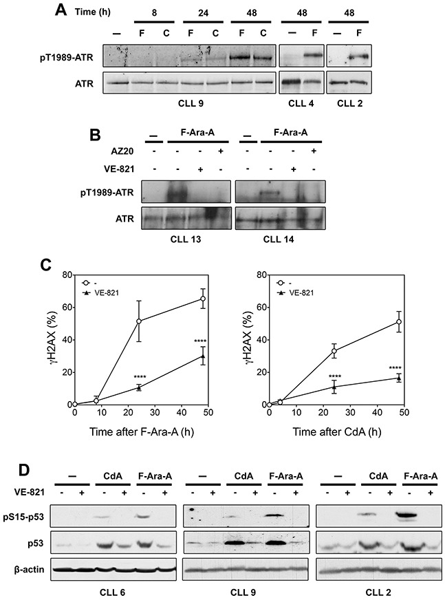
ATM, primarily activated by DNA double-strand breaks, and ATR, activated by single-stranded DNA, are master regulators of the cellular response to DNA damage. In primary chronic lymphocytic leukemia (CLL) cells, ATR signaling is considered to be switched off due to ATR downregulation. Here, we hypothesized that ATR, though expressed at low protein level, could play a role in primary resting CLL cells after genotoxic stress. By investigating the response of CLL cells to UV-C irradiation, a prototypical activator of ATR, we could detect phosphorylation of ATR at Thr-1989, a marker for ATR activation, and also observed that selective ATR inhibitors markedly decreased UV-C-induced phosphorylation of ATR targets, including H2AX and p53. Similar results were obtained with the purine analogs fludarabine and cladribine that were also shown to activate ATR and induce ATR-dependent phosphorylation of H2AX and p53. In addition, ATR inhibition was found to sensitize primary CLL cells to UV-C by decreasing DNA repair synthesis. Conversely, ATR inhibition rescued CLL cells against purine analogs by reducing expression of the pro-apoptotic genes PUMA and BAX. Collectively, our study indicates that ATR signaling can be activated in resting CLL cells and play a pro-survival or pro-apoptotic role, depending on the genotoxic context.

Keywords: ATR; CLL; UV-C; fludarabine; p53.


Conflict of interest statement

CONFLICTS OF INTEREST The authors declare no conflicts of interest.

Figures

Figure 1
Figure 1. ATR protein level in primary resting CLL cells as compared with proliferating cells
Same amounts of lysates obtained from different CLL samples or from EHEB and GM0536 cells were analyzed by immunoblotting for ATR, using a Santa Cruz (A) or a Cell Signaling antibody (B). Cyclin A (C) was analyzed to verify the quiescent state of CLL cells. β-Actin was used as a loading control.
Figure 2
Figure 2. Analysis of ATR signaling in primary resting CLL cells after UV-C irradiation
(A) Time- and dose-dependent activation of ATR by UV-C. CLL cells from 2 different patients were irradiated with UV-C (18 J/m2) and incubated for the indicated times (upper panel), or irradiated with increasing UV-C doses and incubated for 5 h (lower panel). Cell lysates were probed for phospho-ATR(Thr-1989) and ATR protein level. (B) Effect of ATR and ATM inhibitors on UV-C-induced γH2AX accumulation. CLL cells were preincubated with or without 10 μM VE-821, 1 μM AZ20 or 10 μM KU-60019 before UV-C irradiation (18 J/m2). At the indicated times, phosphorylation of H2AX (γH2AX) was analyzed by flow cytometry and expressed as fold increase over untreated cells. Results are means ± SEM of 5 independent experiments. (C) Time- and dose-dependent activation of p53 after UV-C exposure. CLL cells were irradiated with UV-C (18 J/m2) and incubated for the indicated times (upper panel), or irradiated with UV-C at 12 and 18 J/m2 and incubated for 5 h (lower panel). Cell lysates were probed for phospho-p53(Ser-15) and p53 protein level. GAPDH or β-actin were used as loading controls. (D) Effect of VE-821 on UV-C-induced p53 activation. CLL cells from two different patients were preincubated with 10 μM VE-821 before UV-C irradiation (18 J/m2). Phosphorylation of p53 at Ser-15 and p53 protein level were analyzed by Western blot 5 h (upper panel) or 8 h (lower panel) after irradiation. (E) Effect of ATR inhibitors on dCK activation and phosphorylation induced by UV-C. CLL cells were preincubated with or without 10 μM VE-821 or 1 μM AZ20 before UV-C irradiation (18 J/m2). dCK activity (n = 3, left panel) and phospho-dCK(Ser-74) (right panel) were analyzed 30 min after irradiation. Basal dCK activity was 10.1 ± 1.7 pmol/min/mg protein (n = 3). Significance relative to the absence of ATR inhibitors (panels B and E): ****P < 0.0001.
Figure 3
Figure 3. Analysis of ATR signaling in primary resting CLL cells after treatment with fludarabine (F-Ara-A) or cladribine (CdA)
(A) Activation of ATR by purine analogs. CLL cells were incubated with 3 μM F-Ara-A (F) or 1 μM CdA (C) for various times, or with or without 3 μM F-Ara-A for 48 h. Phospho-ATR(Thr-1989) and ATR protein level were analyzed by Western blot. (B) Effect of ATR inhibitors on fludarabine-induced ATR phosphorylation. CLL cells were incubated for 48 h with or without 3 μM F-Ara-A in the presence or absence of 10 μM VE-821 or 1 μM AZ20, followed by analysis of ATR phosphorylation by Western blot. (C) Effect of VE-821 on γH2AX accumulation induced by F-Ara-A (left panel) or CdA (right panel). CLL cells were preincubated with or without 10 μM VE-821 before addition of 3 μM F-Ara-A or 1 μM CdA. At the indicated times, phosphorylation of H2AX (γH2AX) was analyzed by flow cytometry and expressed as fold-increase over untreated cells. Results are means ± SEM of 4 independent experiments. Significance relative to the absence of VE-821: ****P < 0.0001. (D) Effect of VE-821 on p53 activation induced by fludarabine or cladribine. CLL cells from three different patients were preincubated with 10 μM VE-821 before addition of 1 μM CdA or 3 μM F-Ara-A. Phospho-p53(Ser-15) and p53 protein level were analyzed by Western blot after 24 h of incubation. β-Actin were used as a loading control.
Figure 4
Figure 4. Effect of ATR inhibitors on cell death induced by UV-C irradiation or purine analogs in primary resting CLL cells
(A) Effect of VE-821 on UV-C-induced cytoxicity. CLL cells were preincubated with or without 10 μM VE-821 and then exposed to increasing UV-C doses, as indicated. Cell viability was measured by the MTT assay after 24 h of incubation. Results are means ± SEM of 4 separate experiments. (B) Effect of VE-821 on caspase-3 activation induced by UV-C irradiation. CLL cells were preincubated with or without 10 μM VE-821 and then irradiated with UV-C at 12 or 18 J/m2. Caspase-3 activity was analyzed 8 h after irradiation by an enzymatic assay (n=3, upper panel) or by immunoblotting of its active cleaved form (lower panel). (C-D) Influence of ATR inhibitors on purine analog-induced cytotoxicity. CLL cells were incubated for 48 h with F-Ara-A (C) or CdA (D) at increasing concentrations in the absence or presence of VE-821 or AZ20. Cell viability was measured by the MTT assay. Results are means ± SEM of 4 separate experiments. (E) Effect of AZD6738 as compared to VE-821 on F-Ara-A-induced cytotoxicity. CLL cells were incubated for 96 h with F-Ara-A at increasing concentrations in the presence or absence of 10 μM VE-821 or 1 μM AZD6738. Cell viability was measured by the MTT assay. Results are means ± SEM of 3 separate experiments. (F) Effect of VE-821 on caspase-3 activation induced by CdA or F-Ara-A. CLL cells were preincubated with or without 10 μM VE-821 and incubated in the presence or absence of 1 μM CdA or 3 μM F-Ara-A. Caspase-3 activity was analyzed after 48 h by an enzymatic assay (n = 3, upper panel) or by immunoblotting of its active cleaved form (lower panel). (G) Effect of VE-821 when added 24 h after fludarabine. CLL cells were incubated for 24 h in the presence or absence of 3 μM F-Ara-A, washed from F-Ara-A, and resuspended in fresh medium containing, or not, 10 μM VE-821. Caspase-3 activity was measured just before washing and after additional 24 h, by an enzymatic assay (upper panel). Results are means ± SEM of 4 independent experiments. p53 phosphorylation and p53 protein level were also analyzed at the same incubation times by Western blot (lower panel). GAPDH was used as a loading control. The dotted lines in the upper and lower panels indicate washing of cells to remove F-Ara-A. Significance relative to the absence of ATR inhibitors: *P < 0.05; **P <0.01; ***P < 0.001; ****P < 0.0001.
Figure 5
Figure 5. Effect of ATR inhibition on DNA repair synthesis and pro-apoptotic gene expression in primary resting CLL cells after treatment with UV-C or purine analogs
(A) DNA repair synthesis after UV-C irradiation. After preincubation with or without 10 μM VE-821, CLL cells were, or not, UV-C-irradiated (18 J/M2) and incubated with labeled thymidine (Thd) for 3 h. Results are means ± SEM of 3 independent experiments. (B) DNA repair synthesis after treatment with F-Ara-A. After preincubation with or without 10 μM VE-821, CLL cells were incubated in the absence or presence of 3 μM F-Ara-A for 43 h, and then incubated with labeled thymidine for 5 h. Results are means ± SEM of 3 independent experiments. (C) PUMA and BAX expression after UV-C irradiation. After preincubation with or without 10 μM VE-821, CLL cells (n = 3) were UV-C irradiated (18 J/m2) and incubated for 8 h before analysis of PUMA and BAX expression by qPCR (upper and middle panels) and Western blot (lower panel). (D) PUMA and BAX expression after purine analogs. After preincubation with or without 10 μM VE-821, CLL cells (n = 3) were incubated with 1 μM CdA or 3 μM F-Ara-A for 24 h before analysis of PUMA and BAX expression by qPCR (upper and middle panel) and Western blot (lower panel). Significance relative to the absence of VE-821 (panels A, B and D): *P < 0.05; **P <0.01.
Figure 6
Figure 6. Effect of ATR inhibition on fludarabine- and UV-C -induced cytotoxicity in p53-defective CLL cells
(A) 17p-deleted CLL samples (CLL12, 23 and 40), containing ≥ 71 % of cells with deletion, were incubated for 96 h with increasing concentration of F-Ara-A in the presence or absence of 10 μM VE-821. (B) One of these samples (CLL12) was also irradiated with increasing UV-C doses and incubated for 24 h in the presence or absence of 10 μM VE-821. Cell survival was measured by the MTT assay.

References

    1. Chiorazzi N, Rai KR, Ferrarini M. Chronic lymphocytic leukemia. N Engl J Med. 2005;352:804–815. - PubMed
    1. Cramer P, Hallek M. Prognostic factors in chronic lymphocytic leukemia-what do we need to know? Nat Rev Clin Oncol. 2011;8:38–47. - PubMed
    1. Dohner H, Fischer K, Bentz M, Hansen K, Benner A, Cabot G, Diehl D, Schlenk R, Coy J, Stilgenbauer S, Volkmann M, Galle PR, Poustka A, et al. p53 gene deletion predicts for poor survival and non-response to therapy with purine analogs in chronic B-cell leukemias. Blood. 1995;85:1580–1589. - PubMed
    1. Zenz T, Eichhorst B, Busch R, Denzel T, Habe S, Winkler D, Buhler A, Edelmann J, Bergmann M, Hopfinger G, Hensel M, Hallek M, Dohner H, et al. TP53 mutation and survival in chronic lymphocytic leukemia. J Clin Oncol. 2010;28:4473–4479. - PubMed
    1. Roos WP, Kaina B. DNA damage-induced cell death: from specific DNA lesions to the DNA damage response and apoptosis. Cancer Lett. 2013;332:237–248. - PubMed

LinkOut - more resources