Review
. 2017 May 23;8(34):57800-57812.
doi: 10.18632/oncotarget.18101. eCollection 2017 Aug 22.

Formation, function, and exhaustion of notochordal cytoplasmic vacuoles within intervertebral disc: current understanding and speculation

Affiliations
Review

Formation, function, and exhaustion of notochordal cytoplasmic vacuoles within intervertebral disc: current understanding and speculation

Feng Wang et al. Oncotarget. .

Abstract

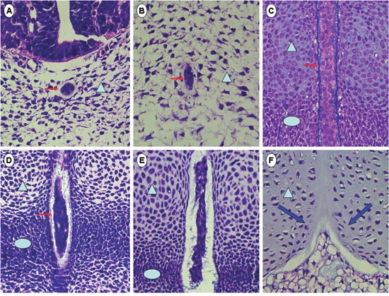
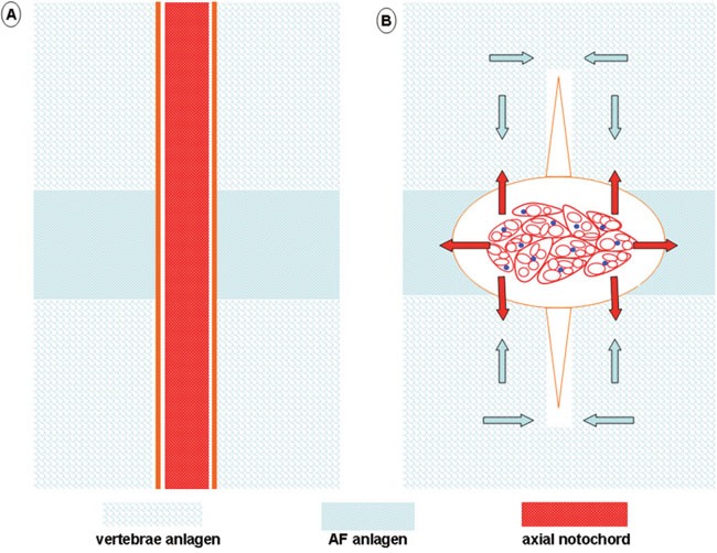
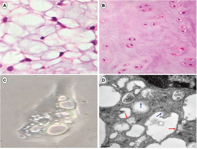
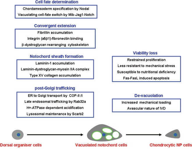
Notochord nucleus pulposus cells are characteristic of containing abundant and giant cytoplasmic vacuoles. This review explores the embryonic formation, biological function, and postnatal exhaustion of notochord vacuoles, aiming to characterize the signal network transforming the vacuolated nucleus pulposus cells into the vacuole-less chondrocytic cells. Embryonically, the cytoplasmic vacuoles within vertebrate notochord originate from an evolutionarily conserved vacuolation process during neurulation, which may continue to provide mechanical and signal support in constructing a mammalian intervertebral disc. For full vacuolation, a vacuolating specification from dorsal organizer cells, synchronized convergent extension, well-structured notochord sheath, and sufficient post-Golgi trafficking in notochord cells are required. Postnatally, age-related and species-specific exhaustion of vacuolated nucleus pulposus cells could be potentiated by Fas- and Fas ligand-induced apoptosis, intolerance to mechanical stress and nutrient deficiency, vacuole-mediated proliferation check, and gradual de-vacuolation within the avascular and compression-loaded intervertebral disc. These results suggest that the notochord vacuoles are active and versatile organelles for both embryonic notochord and postnatal nucleus pulposus, and may provide novel information on intervertebral disc degeneration to guide cell-based regeneration.

Keywords: cytoplasmic vacuole; intervertebral disc; notochord; notochord vacuolation; nucleus pulposus.


Conflict of interest statement

CONFLICTS OF INTEREST None of the authors have any conflicts of interest to declare.

Figures

Figure 1
Figure 1. Hematoxylin and eosin staining of rat axial notochord from embryonic day 10 (E10) to E18.5
As early as E10 ((A), transverse section; (B), coronal section), the axial notochord (arrow) had separated from paraxial mesoderm (arrowhead), although the peri-notochord basement membrane had not fully formed. At E12 (C), the axial notochord was enwrapped by a thick fibrotic sheath (arrow), lateral to which the paraxial mesoderm had patterned into non-condensed sclerotome (arrowhead) and condensed sclerotome (ellipse). From E13.5 (D) to E14.5 (E), the notochord sheath became thinner as the non-condensed sclerotome transformed into vertebrae anlagen (arrowhead) and the condensed sclerotome into AF anlagen (ellipse), although the notochord cells had not been vacuolated at this time. The gap between axial notochord and paraxial mesoderm was widened during this stage (E13.5–14.5), which was probably caused by convergent extension and/or increased intra-notochord pressure induced by high secretion from notochord cells. From E14.5 to E18.5 (F), the paired non-condensed sclerotome fused symmetrically (blue arrow) to form the vertebral body (arrowhead), whereas the notochord cells vacuolated and were squeezed into the center of IVD, probably by the pushing pressure generated from the forming vertebrae. Original magnification ×400.
Figure 2
Figure 2. Schematic illustration of IVD formation and mechanical support from notochord vacuolation
During the formation of mammalian IVD, the paired vertebrae anlagen (non-condensed sclerotome) fuses and compresses the axial notochord into the center of forming IVD, the paired AF anlagen (condensed sclerotome) expands outward and enwraps the vacuolating notochord. By generating even resistance (red arrow) to the paired inward-expanding (blue arrow) vertebral anlagen, the symmetrical morphology of vertebral body could be maintained by the vacuolating notochord. By producing a primitive swelling tension (red arrow) inside the forming IVD, the vacuolating notochord might also contribute to condense the forming AF and maintain IVD height between the growing vertebra.
Figure 3
Figure 3. Exhaustion of notochord nucleus pulposus cells
Hematoxylin and eosin staining of neonatal (A) and aged (B) rat NP showed the replacement of vacuolated notochord NP cells (NNPCs) by non-vacuolated chondrocytic NP cells (CNPCs) and fibrocartilaginous extracellular matrix during natural aging. In monolayer cultures (C), the NNPCs were rich in giant cytoplasmic vacuoles. On transmission electron micrographs (D), the vacuole membrane (red arrow) in NNPCs isolated from adult rat was partially or completely lost (blue arrow), suggesting that a de-vacuolation process may also contribute to the loss of NNPCs. Original magnification ×400 for (A–C).
Figure 4
Figure 4. Summary of the signal basis underlying the generation and exhaustion of notochord vacuoles
Notochord vacuolation is a highly coordinated and orchestrated process that requires vacuolating specification from dorsal organizer cells, intact convergent extension, formation of notochord sheath, as well as sufficient post-Golgi trafficking within the notochord cells themselves. After squeezing into the IVD, the vacuolated notochord NP cells can be exhausted by inferior cell viability and a chronic process of de-vacuolation, which may result from the increased mechanical stress and avascular nature of postnatal IVD.

Similar articles

Cited by

References

    1. Vos T, Flaxman AD, Naghavi M, Lozano R, Michaud C, Ezzati M, Shibuya K, Salomon JA, Abdalla S, Aboyans V, Abraham J, Ackerman I, Aggarwal R, et al. Years lived with disability (YLDs) for 1160 sequelae of 289 diseases and injuries 1990-2010: a systematic analysis for the Global Burden of Disease Study 2010. Lancet. 2012;380:2163–96. - PMC - PubMed
    1. Hoy D, Bain C, Williams G, March L, Brooks P, Blyth F, Woolf A, Vos T, Buchbinder R. A systematic review of the global prevalence of low back pain. Arthritis Rheum. 2012;64:2028–37. - PubMed
    1. Risbud MV, Shapiro IM. Role of cytokines in intervertebral disc degeneration: pain and disc content. Nat Rev Rheumatol. 2014;10:44–56. - PMC - PubMed
    1. Huang YC, Urban JP, Luk KD. Intervertebral disc regeneration: do nutrients lead the way. Nat Rev Rheumatol. 2014;10:561–6. - PubMed
    1. Wang F, Cai F, Shi R, Wang XH, Wu XT. Aging and age related stresses: a senescence mechanism of intervertebral disc degeneration. Osteoarthritis Cartilage. 2016;24:398–408. - PubMed