. 2017 Sep 15;17(1):217.
doi: 10.1186/s12862-017-1057-1.

Morphogenesis along the animal-vegetal axis: fates of primary quartet micromere daughters in the gastropod Crepidula fornicata

Affiliations

Morphogenesis along the animal-vegetal axis: fates of primary quartet micromere daughters in the gastropod Crepidula fornicata

Deirdre C Lyons et al. BMC Evol Biol. .

Abstract

Background: The Spiralia are a large, morphologically diverse group of protostomes (e.g. molluscs, annelids, nemerteans) that share a homologous mode of early development called spiral cleavage. One of the most highly-conserved features of spiralian development is the contribution of the primary quartet cells, 1a-1d, to the anterior region of the embryo (including the brain, eyes, and the anterior ciliary band, called the prototroch). Yet, very few studies have analyzed the ultimate fates of primary quartet sub-lineages, or examined the morphogenetic events that take place in the anterior region of the embryo.

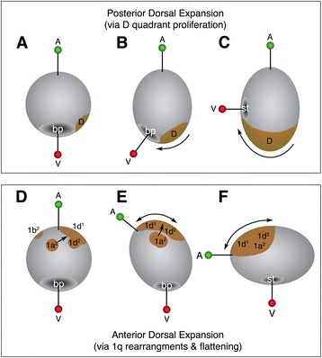
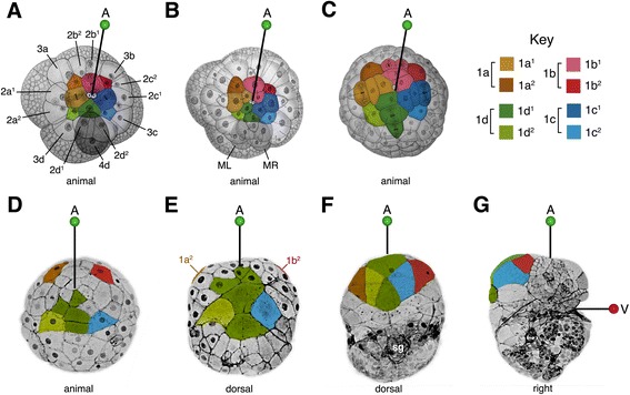
Results: This study focuses on the caenogastropod slipper snail, Crepidula fornicata, a model for molluscan developmental biology. Through direct lineage tracing of primary quartet daughter cells, and examination of these cells during gastrulation and organogenesis stages, we uncovered behaviors never described before in a spiralian. For the first time, we show that the 1a2-1d2 cells do not contribute to the prototroch (as they do in other species) and are ultimately lost before hatching. During gastrulation and anterior-posterior axial elongation stages, these cells cleavage-arrest and spread dramatically, contributing to a thin provisional epidermis on the dorsal side of the embryo. This spreading is coupled with the displacement of the animal pole, and other pretrochal cells, closer to the ventrally-positioned mouth, and the vegetal pole.

Conclusions: This is the first study to document the behavior and fate of primary quartet sub-lineages among molluscs. We speculate that the function of 1a2-1d2 cells (in addition to two cells derived from 1d12, and the 2b lineage) is to serve as a provisional epithelium that allows for anterior displacement of the other progeny of the primary quartet towards the anterior-ventral side of the embryo. These data support a new and novel mechanism for axial bending, distinct from canonical models in which axial bending is suggested to be driven primarily by differential proliferation of posterior dorsal cells. These data suggest also that examining sub-lineages in other spiralians will reveal greater variation than previously assumed.

Keywords: Axial elongation; Gastropoda; Lophotrochozoa; Mollusca; Morphogenesis; Prototroch; Spiralia; Trochoblasts.


Conflict of interest statement

Ethics approval and consent to participate

This study used wild-caught invertebrate snails and those adult animals were all returned back to the ocean after their embryos were collected.

Consent for publication

Not applicable.

Competing interests

The authors declare that they have no competing interests.

Publisher’s Note

Springer Nature remains neutral with regard to jurisdictional claims in published maps and institutional affiliations.

Figures

Fig. 1
Fig. 1
Proposed models describing morphogenetic events that contribute to the bending of the animal-vegetal axis of the spiralian embryo. These events reposition the mouth to the future ventral side of the embryo and closer the animal (future anterior) pole. a-c Earlier model suggesting that the vegetal pole and site of gastrulation (where the blastopore and mouth form) is displaced by differential proliferation of post-trochal D quadrant progeny (mainly 2d progeny) on the posterior dorsal side of the embryo [32]. d-f Model proposed here based on data from C. fornicata where axial bending is driven by ventral displacement of the animal pole through rearrangement and flattening of 1q2 (i.e., 1a1-1d1) and 1d1 (i.e., 1d121 and 1d122) progeny. While these two models are not mutually exclusive, the data reported here revealed the latter process in C. fornicata, and indicate that these cells are repositioned towards the dorsal side of the head to form an expansive, provisional epithelium, which is subsequently lost before hatching. Cells are labeled following the nomenclature of Conklin [40], and as described in the text. bp, blastopore; A, animal pole; st, stomodeum; V, vegetal pole
Fig. 2
Fig. 2
Schematic diagram highlighting cells/clones derived from the first quartet micromeres 1a1-1d1 and 1a2-1d2. These cells/clones are color coded as indicated in the key. a-c Animal pole views with the d quadrant located towards the bottom of the figure. The other micromere progeny are labeled in (a) for reference. The central small round polar bodies (gray color) are located at the animal pole. Note that the 1q2 cells do not undergo subsequent divisions and initially present a very small exposed area on the surface of the embryos, but eventually they spread out to occupy a greater exposed surface area (also see Fig. 3). d-g Diagrams showing later stages and differential movement of the 1q2 micromeres to form the provisional epithelium, with progeny of 1d1, on the dorsal side of the head. Color has been removed from the 1q1 clones for better clarity. These rearrangements help accommodate the relative displacement of the animal pole towards the vegetal/ventral pole during development. The posterior pole is located towards the bottom of the figure for embryos shown in (d-g). d Dorsal view during late cleavage in an embryo undergoing compaction. e Dorsal view of an embryo beginning elongation. f Dorsal view of an older embryo that is beginning organogenesis. The circular, condensed shell gland (sg) has begun to form in this embryo. g Right lateral view of an embryo that is somewhat older than that in (f). Note the animal pole ends up as the anterior pole of the embryo/larva while the vegetal pole (the site of gastrulation, blastopore, and mouth) ends up on the ventral surface. The originally straight animal-vegetal axis becomes bent, and the animal pole becomes located at 90 degrees relative to the vegetal/ventral pole. For each embryo, the animal/anterior pole (A) is indicated with a green-headed pin. The vegetal/ventral pole (V) is indicated with a red-headed pin in g. Embryos in (a-c) follow illustrations of Conklin [40]. Embryos in (d-g) are from confocal images where the cell outlines are visualized by expression of GFP-tagged UTPH (from [29])
Fig. 3
Fig. 3
Cell lineage fate map for C. fornicata, summarizing the results presented here for the first quartet progeny (see also [37]). Nomenclature follows that of Conklin [40]. Fates shown in italics highlight information gleaned from the present study
Fig. 4
Fig. 4
Live images showing examples of specific clones at various stages of development, as labeled. Individual cells were initially injected at the 16- to 28-cell stages. a Three examples of freshly injected 1q2 cells prior to their division (i.e., 1a2 and 1d2 at 28-cell stage, as labeled). Note the small apparent size of these micromere daughters. Nuclei were pre-labeled by expression of green GFP-histone H2B. Red fluorescent diI has been pressure microinjected into single cells using a fine glass needle, as shown. b-f 1a1 and 1a2 progeny, as labeled. b, c Dorsal-animal views of elongating embryos. d, e Left-lateral views of pre-veliger larvae undergoing organogenesis. f Anterior-dorsal view of an early veliger larvae. g-l 1b1 and 1b2 progeny, as labeled. g Animal view with the d quadrant located towards the bottom of the figure. h Dorsal-animal view. i Ventral view. j, k Right lateral views of pre-veliger larvae undergoing organogenesis. l Right lateral view of veliger larva. m-r 1c1 and 1c2 progeny, as labeled. m Animal pole view. n Dorsal-animal view. o Dorsal view. p-q Right-lateral views of pre-veliger larvae undergoing organogenesis. r Anterior-dorsal view of an early veliger larvae. s-x 1d1 and 1d2 progeny, as labeled. s-t Dorsal views of elongating embryos. u-v Right- and left-lateral views, respectively, of pre-veliger larvae undergoing organogenesis. w-x Anterior-dorsal and right-lateral views of early veliger larvae, respectively. To facilitate the injections embryos were pre-labeled by expression of either green GFP-histone H2B (as shown in a, g, h, n, q) or GFP-UTPH (as shown in b, c, i, m, s, t, v) protein. Unless noted otherwise, embryos are oriented with the posterior pole directed toward the bottom of the figure. Scale bar in X equals 50 μm
Fig. 5
Fig. 5
Anti-acetylated tubulin staining (red fluorescence) revealing ciliated cells in developing embryos. Embryos are oriented with the future posterior pole toward the bottom of the figure. Nuclei are labeled with DAPI (blue fluorescence). Specific cells/clones are as labeled. a, b Animal and vegetal views during early gastrulation, respectively. No ciliation is present at this stage. c, d Animal and vegetal views, respectively, during intermediate stages of gastrulation when ciliation is present in some 1q2 daughter cells, as labeled. e, f Animal-dorsal, and vegetal views later during gastrulation, respectively. Note group of four large ciliated cells on the dorsal surface in addition to 1a2 and 1b2. g, h Dorsal and ventral views, respectively, at an even later stage of gastrulation. Note even closer location of 1a2 and 1b2 relative to the four large ciliated cells on the dorsal surface (g). I-L Dorsal, right-lateral, ventral and left-lateral views of elongating embryo, respectively. Ciliated cells of the neurotroch (3c2 and 3d2 progeny) are seen within and posterior to the stomodeum [29]. The ciliated 1q2 cells along with 1d121 and 1d122 now occupy a large area on the dorsal-lateral surfaces of the future head. m-p Dorsal, right-lateral, ventral and left-lateral views, respectively, of a later stage when the embryo is undergoing elongation. Note large area occupied by ciliated cells on the dorsal surface of the head that contribute to the “provisional epithelium.” Additional cells have developed ciliation including progeny of 1b1, which will contribute to the anterior head field and prototroch, in addition to some progeny of 2b1 and 2b2. q-t Later stage of development when organ rudiments form. A large area is occupied by the provisional epithelium on the dorsal surface of the head. u-x Very early veliger larval stage. At this stage the ciliated prototroch cells have formed. There are ciliated cells in the head field and along the ventral midline in the foot (derived from 3c2 and 3d2). bp, blastopore; fr, foot rudiment; ft., foot; st, stomodeum; tc, terminal (“anal”) cells; vl, velar lobes; vr, velar rudiment. Scale bar in X equals 50 μm
Fig. 6
Fig. 6
Embryos stained with DAPI to show the distribution of nuclei at four successive stages of development in C. fornicata. The formation of the expansive provisional epithelium, comprised of a small number of cells (nuclei) can be seen in (d, f, and h). a-b Corresponding fluorescence micrographs showing the vegetal (a) and animal (b) sides, respectively in a round stage embryo undergoing early stages of epiboly. The large opening of the blastopore is seen in (a), which is sparsely populated with cells, as the progeny of the animal micromeres have not yet made it to the vegetal side. Notice the fairly symmetrical pattern of the cells (nuclei) in the animal hemisphere in (b). c-d Corresponding fluorescence micrographs showing the vegetal (d) and animal (d) sides, respectively, of a flattened embryo undergoing later stages of epiboly. c The blastopore has closed considerably. d Notice that cells are somewhat more dispersed on the future dorsal side of the embryo as the provisional epithelium is beginning to form. e-f Corresponding fluorescence micrographs showing the ventral and dorsal sides, respectively, of an embryo undergoing elongation. The provisional epithelium is forming on the dorsal surface. g-h Corresponding fluorescence micrographs showing the ventral and dorsal sides, respectively, of an older embryo that has started organogenesis. h The provisional epithelium is present on the dorsal surface of the head, and the shell gland has begun to form in the post-trochal region. Additional nuclei appear to be present in the region of the provisional epithelium, as the deeper endodermal macromeres have undergone some divisions by this stage of development. a, anterior; bp, blastopore; d, dorsal; P, posterior; pe, provisional epithelium; sg, shell gland; st, stomodeum; V, ventral. Asterisk in b, d, e-h marks the location of the animal pole. Scale bar in h equals 50 μm
Fig. 7
Fig. 7
Summary diagram showing behavior of the 1q2 micromeres and 1q1 progeny at later stages of development. Individual cells and their clones are colored, as shown in (a). Orientations or views are indicated under each diagram. Embryos (b-h) are oriented with the future posterior pole directed toward the bottom of the figure. The animal pole is marked with a pink dot in each embryo. a Animal pole view of 25-cell stage and color key. b-e Four different views of an early organogenesis stage embryo showing contributions of the 1q progeny to the developing head. Note that the 1q2 cells now occupy a much smaller area on the dorsal surface of the head. f-h Three views of an advanced veliger larva showing contributions of the formation of the head, anterior surface of the velum and the prototroch by progeny of the 1q1 cells. By this stage the 1q2 cells have been lost from the embryo. ft., foot; lvl, left velar lobe; rvl, right velar lobe; sh, shell; st, stomodeum. Based on the result of this study, as well as those of Hejnol et al. [37]

References

    1. Goldstein B, Freeman G. Axis specification in animal development. BioEssays. 1997;19:105–16. - PubMed
    1. Martindale MQ. The evolution of metazoan axial properties. Nat Rev Genet. 2005;6:917–927. doi: 10.1038/nrg1725. - DOI - PubMed
    1. Hejnol A, Martin-Durán JM. Getting to the bottom of anal evolution. Zoologischer Anzeiger - A J Comp Zool. 2015;256:61–74. doi: 10.1016/j.jcz.2015.02.006. - DOI
    1. Martindale MQ, Hejnol A. A developmental perspective: changes in the position of the blastopore during bilaterian evolution. Dev Cell. 2009;17:162–174. doi: 10.1016/j.devcel.2009.07.024. - DOI - PubMed
    1. Munro E, Robin F, Lemaire P. Cellular morphogenesis in ascidians: how to shape a simple tadpole. Curr Opin Genet Dev. 2006;16:399–405. doi: 10.1016/j.gde.2006.06.004. - DOI - PubMed