. 2017 Sep 15;19(1):203.
doi: 10.1186/s13075-017-1418-6.

MicroRNA-488 and -920 regulate the production of proinflammatory cytokines in acute gouty arthritis

Affiliations

MicroRNA-488 and -920 regulate the production of proinflammatory cytokines in acute gouty arthritis

Weidong Zhou et al. Arthritis Res Ther. .

Abstract

Background: Gout is considered one of the most painful acute conditions caused by deposition of monosodium urate (MSU) crystals within joints. Recent studies have shown that interleukin (IL)-1β is a key inflammatory mediator in acute gouty arthritis (GA), and its level is regulated by microRNAs (miRNAs). However, the molecular mechanisms of the regulation remain unclear.

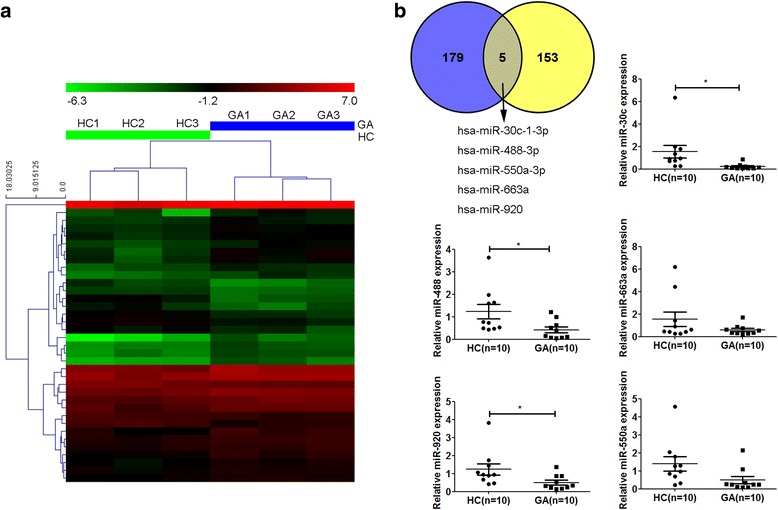
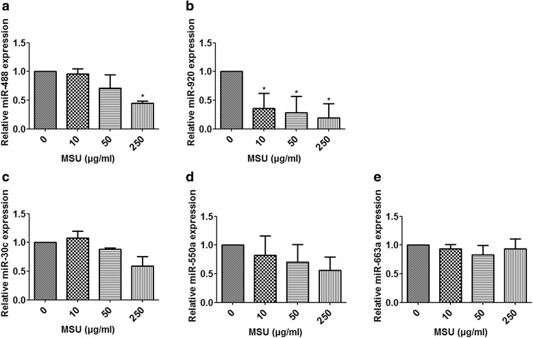
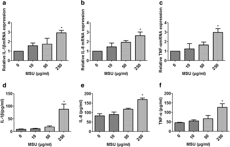
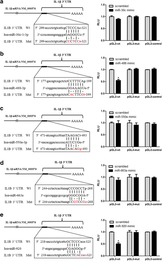
Methods: A miRNA microarray was used to analyze the miRNA expression profiles in peripheral white blood cells (WBCs) of patients with GA. THP-1 cells were transfected with miRNA mimics, stimulated by MSU crystals, and then subjected to quantitative real-time polymerase chain reaction or Western blot analysis. Levels of IL-1β, IL-8, and tumor necrosis factor (TNF)-α in culture supernatants of THP-1 cells were measured by enzyme-linked immunosorbent assay. A luciferase reporter assay was conducted to confirm the interaction of miRNA and IL-1β 3'-untranslated regions (UTRs).

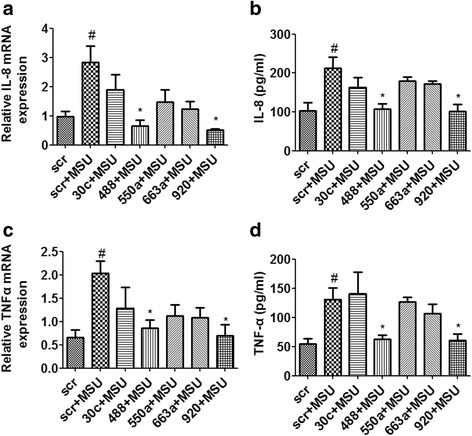
Results: Combining bioinformatics and miRNA expression profiles, we found five miRNAs (hsa-miR-30c-1-3p, hsa-miR-488-3p, hsa-miR-550a-3p, hsa-miR-663a, and hsa-miR-920) that possibly target IL-1β. Then, we demonstrated that miR-488 and miR-920 were significantly decreased in the WBCs of patients with GA and that MSU crystals could inhibit expression of miR-488 and miR-920. Upregulation of miR-488 and miR-920 could suppress MSU-induced IL-1β protein expression in THP-1 cells, but no significant difference in IL-1β messenger RNA levels was observed. Moreover, we found that miR-488 and miR-920 could directly target the 3'-UTR of IL-1β. Overexpression of miR-488 and miR-920 could significantly inhibit the gene and protein expression of IL-8 and TNF-α in MSU-induced THP-1 cells.

Conclusions: This study demonstrates the roles of miR-488 and miR-920 in regulating the production of proinflammatory cytokines in the pathogenesis of GA. These findings suggest that miR-488 and miR-920 could serve as potential therapeutic targets in the treatment of GA.

Keywords: Gouty arthritis; Interleukin-1β; MicroRNA; Monosodium urate crystals; Proinflammatory cytokines.


Conflict of interest statement

Authors’ information

Not applicable.

Ethics approval and consent to participate

This study was approved by the Ethics Committee of the First Affiliated Hospital of Xiamen University. The need for informed consent from the patients was waived.

Consent for publication

Not applicable.

Competing interests

The authors declare that they have no competing interests.

Publisher’s Note

Springer Nature remains neutral with regard to jurisdictional claims in published maps and institutional affiliations.

Figures

Fig. 1
Fig. 1
Identification of microRNAs (miRNAs, miRs) regulating interleukin (IL)-1β expression in human gouty arthritis. a Heat map displays the most differential expression levels in the healthy control subjects (HCs; n = 3) and patients with acute gouty arthritis (GA; n = 3). The color key indicates increasing miRNA expression levels from red to green compared with HC. b Venn diagram (upper left) shows five miRNAs possibly targeting IL-1β in the peripheral white blood cells (WBCs) of patients with GA. Blue represents 184 potential miRNAs targeting human IL-1β gene as predicted by bioinformatics. Yellow represents microarray results of 158 miRNAs markedly downregulated in the WBCs of patients with GA. The expression levels of the five selected miRNAs were detected by quantitative real-time polymerase chain reaction in HCs (n = 10) and patients with GA (n = 10). Values are expressed as mean ± SEM of three independent experiments, each of which was run in triplicate. *P < 0.05
Fig. 2
Fig. 2
Effects of monosodium urate (MSU) crystals on expression of microRNAs (miRNAs, miR) in monocytic THP-1 cells. THP-1 cells were stimulated by the indicated concentration of MSU crystals. The expression levels of miRNAs were detected by quantitative real-time polymerase chain reaction(a-e). Values are expressed as mean ± SEM of three independent experiments, each of which was run in triplicate. *P < 0.05 versus no MSU stimulation
Fig. 3
Fig. 3
Monosodium urate (MSU) crystals promote the expression of proinflammatory cytokines in THP-1 cells. THP-1 cells were stimulated by the indicated concentration of MSU crystals. The messenger (mRNA) expression of interleukin (IL)-1β, IL-8, and tumor necrosis factor (TNF)-α were detected by quantitative real-time polymerase chain reaction (ac). Protein expression of IL-1β, IL-8, and TNF-α was detected by enzyme-linked immunosorbent assay (df). Values are expressed as mean ± SEM of three independent experiments, each of which was run in triplicate. *P < 0.05 versus no MSU stimulation
Fig. 4
Fig. 4
MicroRNA (miR, miRNA)-488 and miR-920 inhibit monosodium urate (MSU)-induced interleukin (IL)-1β protein expression in monocytic THP-1 cells. The miRNA mimics or negative control (NC) mimics (50 nM) were transfected into THP-1 cells using Lipofectamine RNAiMAX reagent in accordance with the manufacturer’s instructions. After 24 h of transfection, cells were stimulated for 3 h with 0.5 μM 12-myristate 13-acetate. Then, cells were washed and stimulated with 250 μg/ml MSU crystals for 24 h to detect the production of IL-1β. After the treatment, the cells were collected and analyzed by quantitative real-time polymerase chain reaction (a) or Western blotting (c). The cell culture supernatants were collected to detect the concentrations of IL-1β by enzyme-linked immunosorbent assay (b). d Densitometric analysis of immunoblot band intensities for IL-1β normalized by β-actin. Values are expressed as mean ± SEM of three independent experiments. # P < 0.05 versus NC, *P < 0.05 versus MSU stimulation alone
Fig. 5
Fig. 5
MicroRNA (miR, miRNA)-488 and miR-920 directly target the 3′-untranslated region (3′-UTR) of interleukin (IL)-1β. Sequence alignments of the miRNA base pair site in the 3′-UTR of IL-1β messenger RNA (mRNA) are shown in the left figures (a-e). The “seed” sequence of miRNA that is complementary to IL-1β is shown in capital letters in the dashed boxes. The mutant sequence (mut) is identical to the wild-type sequence (wt), except the mutated nucleotides are shown in red. Results of luciferase assays with HEK 293 T cells are shown in the right figures (a-e). Cells were cotransfected with wt/mut 3′-UTR with miRNA mimics or negative control as indicated. Twenty-four hours after transfection, luciferase activity was detected using a dual-luciferase reporter assay system according to the manufacturer’s instructions. Values are expressed as mean ± SEM of three independent experiments, each of which was run in triplicate. *P < 0.05 versus scrambled group. RLU Relative light units
Fig. 6
Fig. 6
MicroRNA (miR, miRNA)-488 and miR-920 suppress monosodium urate (MSU)-induced expression of proinflammatory cytokines in THP-1 cells. The miRNA mimics or negative control (NC) mimics (50 nM) were transfected into THP-1 cells using Lipofectamine RNAiMAX reagent in accordance with the manufacturer’s instructions. After 24 h of transfection, cells were stimulated for 3 h with 0.5 μM 12-myristate 13-acetate. Then, cells were washed and stimulated with 250 μg/ml MSU crystals for 24 h to detect the production of proinflammatory cytokines. After the treatment, the cells were collected and analyzed by quantitative real-time polymerase chain reaction (a, c). The cell culture supernatants were also collected to detect the concentrations of interleukin (IL)-8 and tumor necrosis factor (TNF)-α by enzyme-linked immunosorbent assay (b, d). Values are expressed as mean ± SEM of three independent experiments, each of which was run in triplicate. # P < 0.05 versus scrambled (scr) group, *P < 0.05 versus MSU stimulation alone. mRNA Messenger RNA

References

    1. Richette P, Bardin T. Gout Lancet. 2010;375(9711):318–328. doi: 10.1016/S0140-6736(09)60883-7. - DOI - PubMed
    1. Cronstein BN, Terkeltaub R. The inflammatory process of gout and its treatment. Arthritis Res Ther. 2006;8(Suppl 1):S3. doi: 10.1186/ar1908. - DOI - PMC - PubMed
    1. Matsuzaki S, Darcha C. Epithelial to mesenchymal transition-like and mesenchymal to epithelial transition-like processes might be involved in the pathogenesis of pelvic endometriosis. Hum Reprod. 2012;27(3):712–721. doi: 10.1093/humrep/der442. - DOI - PubMed
    1. Martinon F, Petrilli V, Mayor A, Tardivel A, Tschopp J. Gout-associated uric acid crystals activate the NALP3 inflammasome. Nature. 2006;440(7081):237–241. doi: 10.1038/nature04516. - DOI - PubMed
    1. Chen CJ, Shi Y, Hearn A, Fitzgerald K, Golenbock D, Reed G, Akira S, Rock KL. MyD88-dependent IL-1 receptor signaling is essential for gouty inflammation stimulated by monosodium urate crystals. J Clin Invest. 2006;116(8):2262–2271. doi: 10.1172/JCI28075. - DOI - PMC - PubMed

Publication types

LinkOut - more resources