

Optical Imaging Paves the Way for Autophagy Research
- PMID: 28916049
- PMCID: PMC7114199
- DOI: 10.1016/j.tibtech.2017.08.006
Optical Imaging Paves the Way for Autophagy Research
Abstract
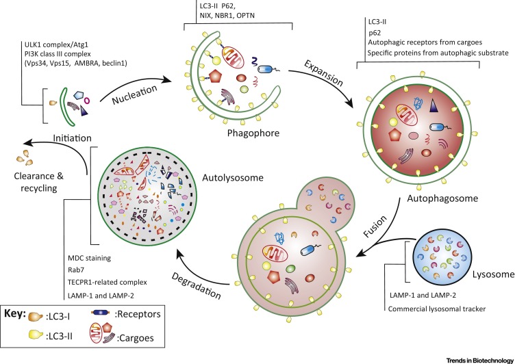
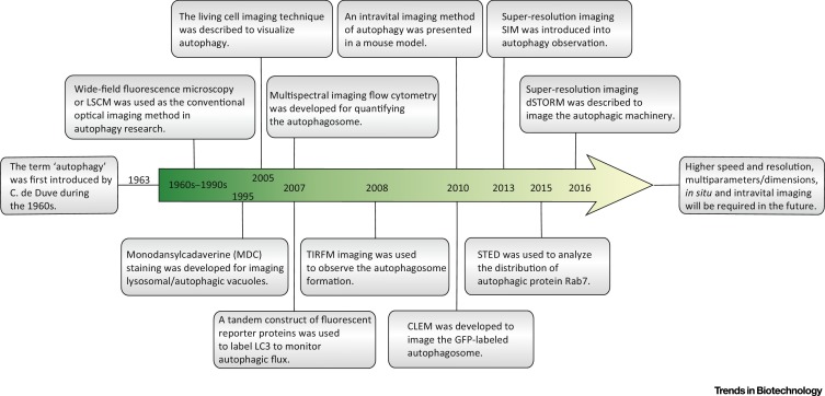
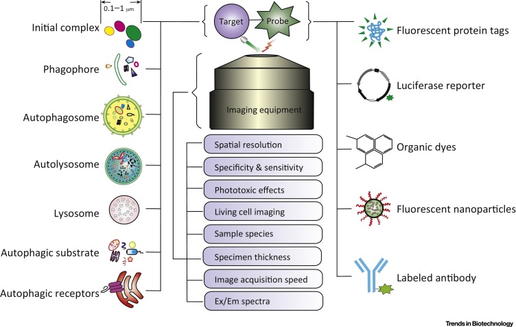
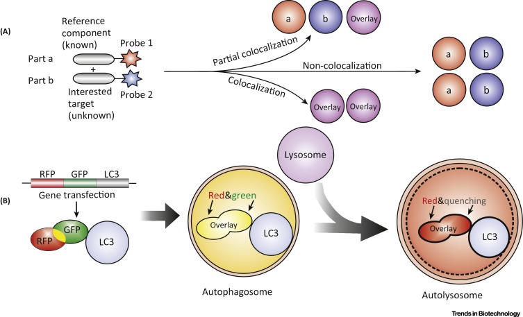
Autophagy is a degradation process in eukaryotic cells that recycles cellular components for nutrition supply under environmental stress and plays a double-edged role in development of major human diseases. Noninvasive optical imaging enables us to clearly visualize various classes of structures involved in autophagy at macroscopic and microscopic dynamic levels. In this review, we discuss important trends of emerging optical imaging technologies used to explore autophagy and provide insights into the mechanistic investigation and structural study of autophagy in mammalian cells. Some exciting new prospects and future research directions regarding optical imaging techniques in this field are also highlighted.
Keywords: autophagy; fluorescent probe; optical imaging; super-resolution.
Copyright © 2017 Elsevier Ltd. All rights reserved.
Figures
Similar articles
-
Fluorescent Bioconjugates for Super-Resolution Optical Nanoscopy.Bioconjug Chem. 2020 Aug 19;31(8):1857-1872. doi: 10.1021/acs.bioconjchem.0c00320. Epub 2020 Jul 24. Bioconjug Chem. 2020. PMID: 32649825
-
Correlative Live-Cell Imaging and Super-Resolution Microscopy of Autophagy.Methods Mol Biol. 2019;1880:231-242. doi: 10.1007/978-1-4939-8873-0_15. Methods Mol Biol. 2019. PMID: 30610701
-
Live Cell Imaging of Mitochondrial Autophagy with a Novel Fluorescent Small Molecule.ACS Chem Biol. 2017 Oct 20;12(10):2546-2551. doi: 10.1021/acschembio.7b00647. Epub 2017 Sep 21. ACS Chem Biol. 2017. PMID: 28925688
-
Technological advances in super-resolution microscopy to study cellular processes.Mol Cell. 2022 Jan 20;82(2):315-332. doi: 10.1016/j.molcel.2021.12.022. Mol Cell. 2022. PMID: 35063099 Free PMC article. Review.
-
State-of-the-art: functional fluorescent probes for bioimaging and pharmacological research.Acta Pharmacol Sin. 2019 Jun;40(6):717-723. doi: 10.1038/s41401-018-0190-8. Epub 2018 Nov 28. Acta Pharmacol Sin. 2019. PMID: 30487651 Free PMC article. Review.
Cited by
-
Quantitative and qualitative analysis of autophagy flux using imaging.BMB Rep. 2020 May;53(5):241-247. doi: 10.5483/BMBRep.2020.53.5.046. BMB Rep. 2020. PMID: 32317089 Free PMC article. Review.
-
Exploiting Nanomaterial-mediated Autophagy for Cancer Therapy.Small Methods. 2019 Feb 13;3(2):1800365. doi: 10.1002/smtd.201800365. Epub 2018 Nov 15. Small Methods. 2019. PMID: 31355327 Free PMC article.
-
De Novo Design of A Membrane-Anchored Probe for Multidimensional Quantification of Endocytic Dynamics.Adv Healthc Mater. 2022 Apr;11(8):e2102185. doi: 10.1002/adhm.202102185. Epub 2022 Jan 27. Adv Healthc Mater. 2022. PMID: 35032365 Free PMC article.
-
ULK1 forms distinct oligomeric states and nanoscopic structures during autophagy initiation.Sci Adv. 2023 Sep 29;9(39):eadh4094. doi: 10.1126/sciadv.adh4094. Epub 2023 Sep 29. Sci Adv. 2023. PMID: 37774021 Free PMC article.
-
Application of Nanomaterials in Biomedical Imaging and Cancer Therapy.Nanomaterials (Basel). 2020 Aug 29;10(9):1700. doi: 10.3390/nano10091700. Nanomaterials (Basel). 2020. PMID: 32872399 Free PMC article. Review.
References
-
- Farkas D.L. Invention and commercialization in optical bioimaging. Nat. Biotechnol. 2003;21:1269–1271. - PubMed
-
- Luo P.G. Carbon “quantum” dots for optical bioimaging. J. Mater. Chem. B. 2013;1:2116–2127. - PubMed
-
- Tian F. In vivo imaging of autophagy in a mouse stroke model. Autophagy. 2010;6:1107–1114. - PubMed
-
- Walker S.A. Correlative live cell and super resolution imaging of autophagosome formation. Methods Enzymol. 2017;587:1–20. - PubMed
Publication types
MeSH terms
Substances
LinkOut - more resources
Full Text Sources
Other Literature Sources
