

Optical Imaging Paves the Way for Autophagy Research
- PMID: 28916049
- PMCID: PMC7114199
- DOI: 10.1016/j.tibtech.2017.08.006
Optical Imaging Paves the Way for Autophagy Research
Abstract
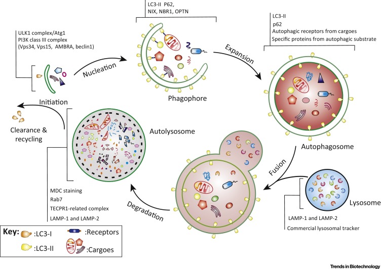
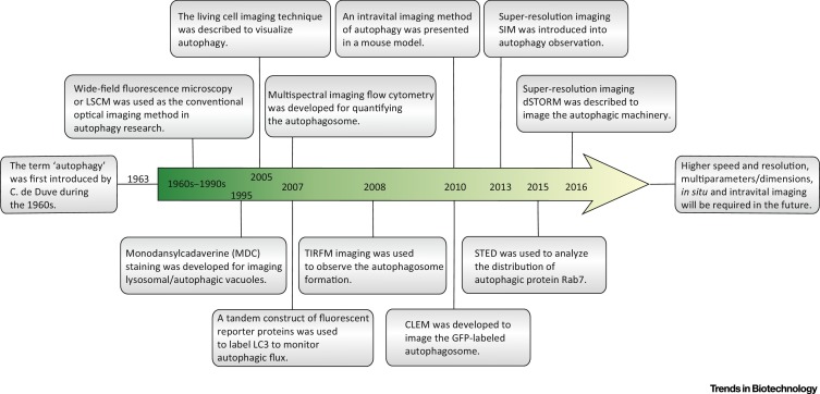
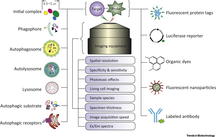
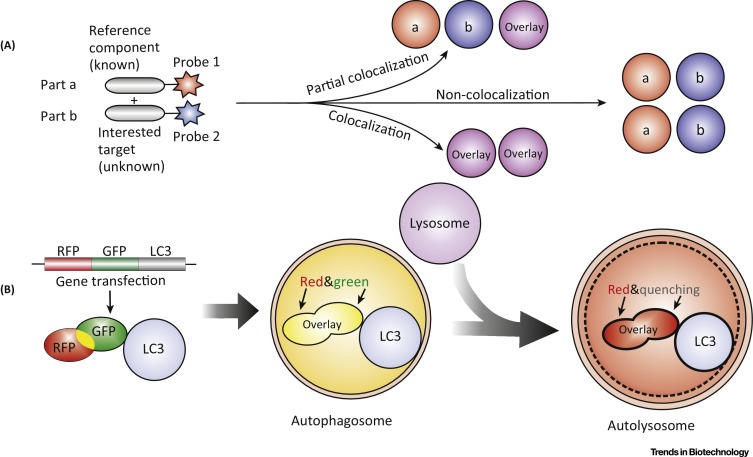
Autophagy is a degradation process in eukaryotic cells that recycles cellular components for nutrition supply under environmental stress and plays a double-edged role in development of major human diseases. Noninvasive optical imaging enables us to clearly visualize various classes of structures involved in autophagy at macroscopic and microscopic dynamic levels. In this review, we discuss important trends of emerging optical imaging technologies used to explore autophagy and provide insights into the mechanistic investigation and structural study of autophagy in mammalian cells. Some exciting new prospects and future research directions regarding optical imaging techniques in this field are also highlighted.
Keywords: autophagy; fluorescent probe; optical imaging; super-resolution.
Copyright © 2017 Elsevier Ltd. All rights reserved.
Figures
References
-
- Farkas D.L. Invention and commercialization in optical bioimaging. Nat. Biotechnol. 2003;21:1269–1271. - PubMed
-
- Luo P.G. Carbon “quantum” dots for optical bioimaging. J. Mater. Chem. B. 2013;1:2116–2127. - PubMed
-
- Tian F. In vivo imaging of autophagy in a mouse stroke model. Autophagy. 2010;6:1107–1114. - PubMed
-
- Walker S.A. Correlative live cell and super resolution imaging of autophagosome formation. Methods Enzymol. 2017;587:1–20. - PubMed
Publication types
MeSH terms
Substances
LinkOut - more resources
Full Text Sources
Other Literature Sources
