. 2017 Sep 19;47(3):498-509.e6.
doi: 10.1016/j.immuni.2017.08.007. Epub 2017 Sep 12.

SIDT2 Transports Extracellular dsRNA into the Cytoplasm for Innate Immune Recognition

Affiliations

SIDT2 Transports Extracellular dsRNA into the Cytoplasm for Innate Immune Recognition

Tan A Nguyen et al. Immunity. .

Abstract

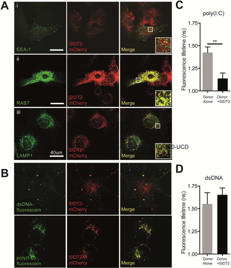
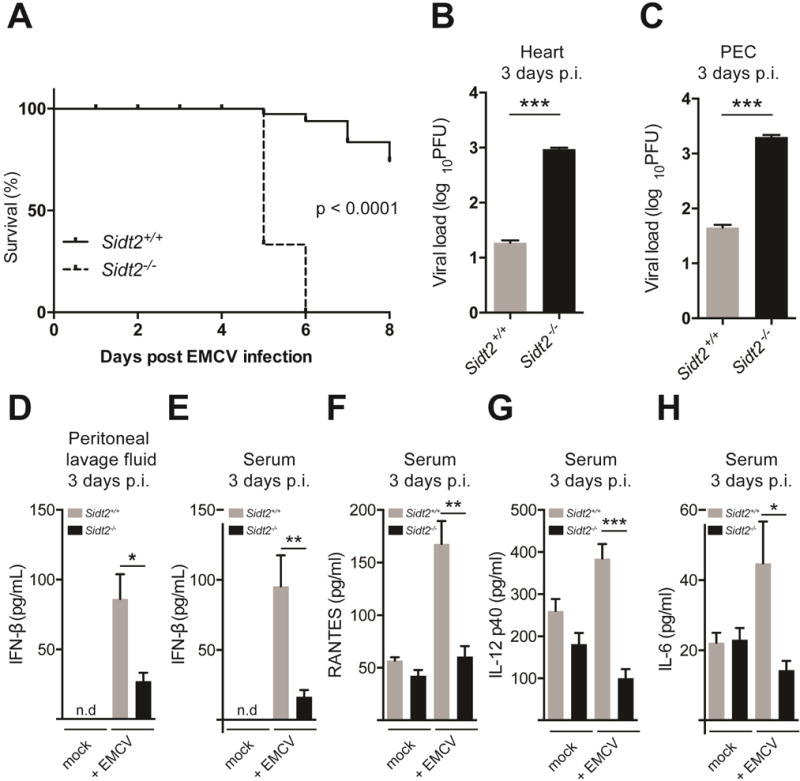
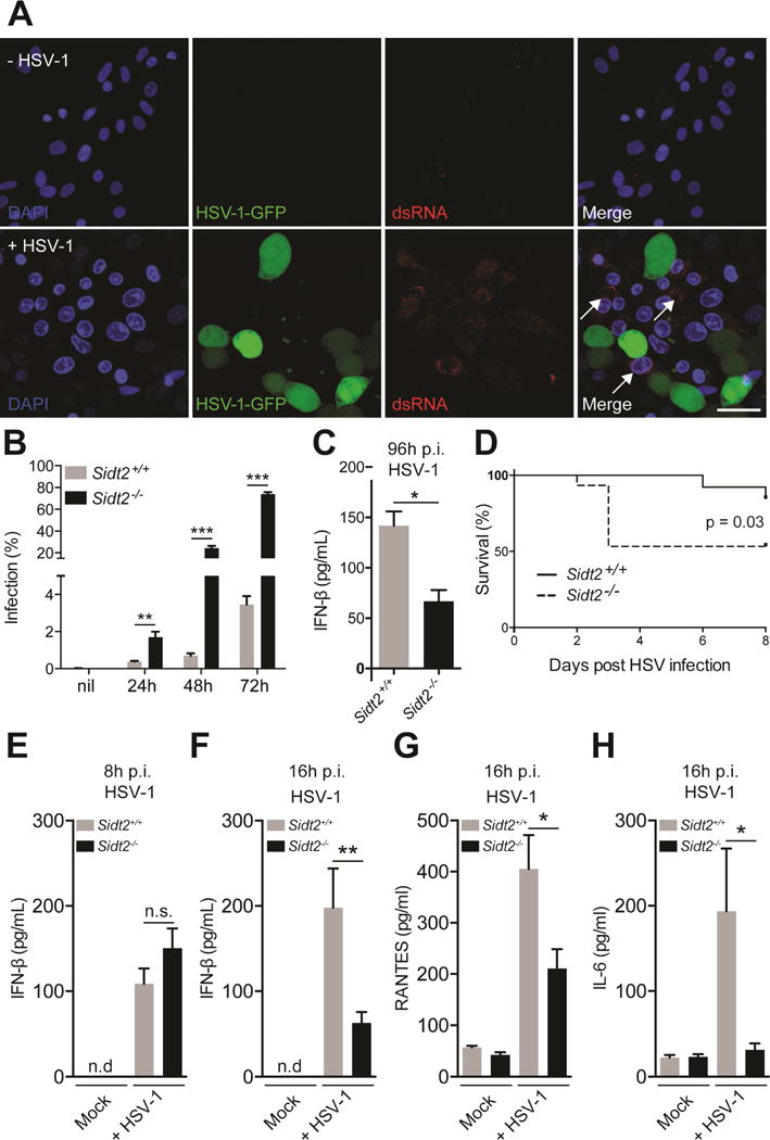
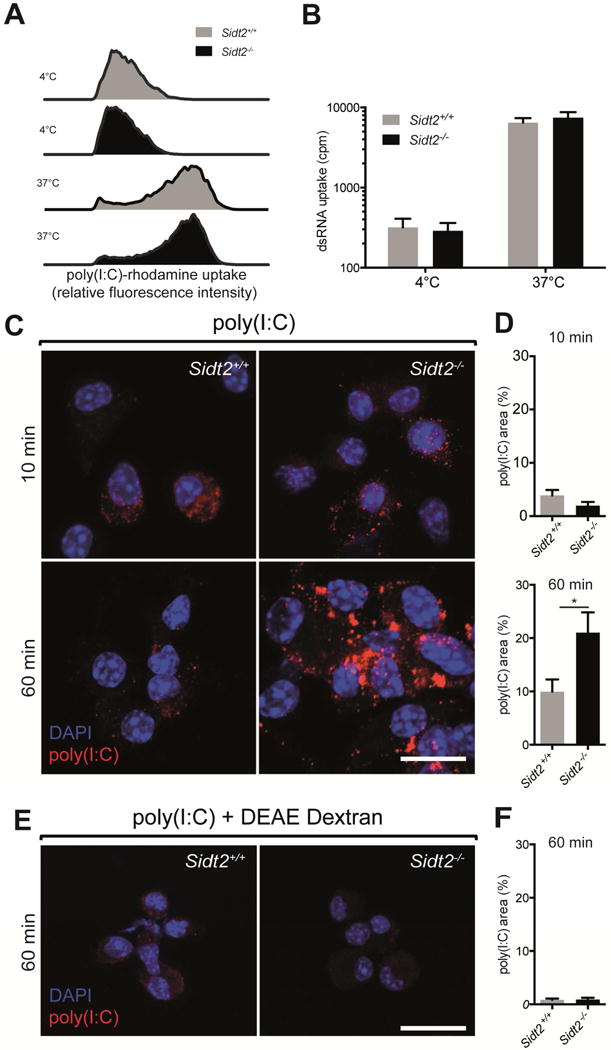
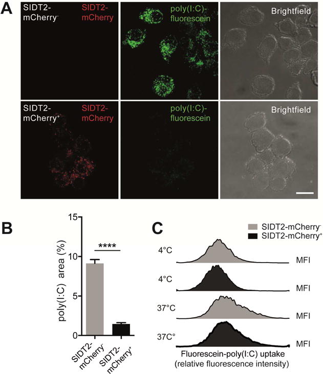
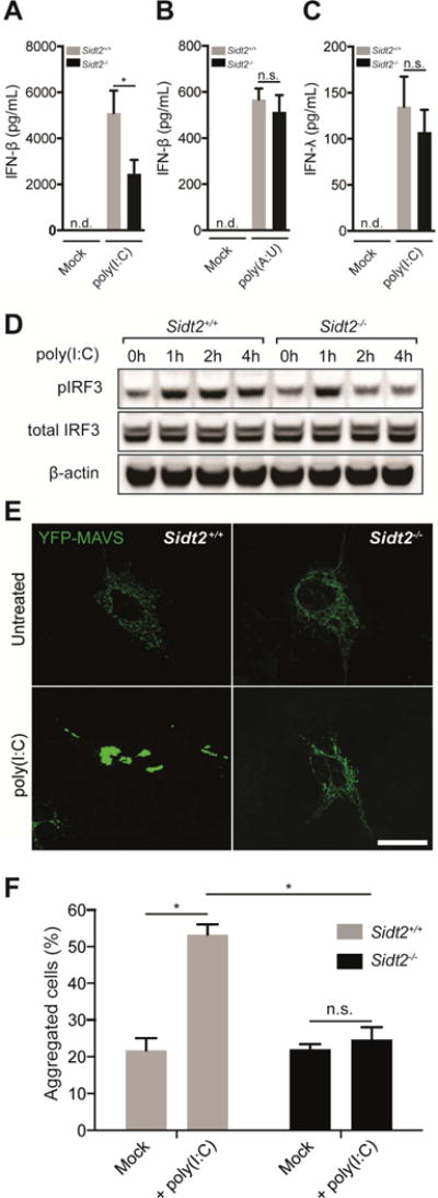
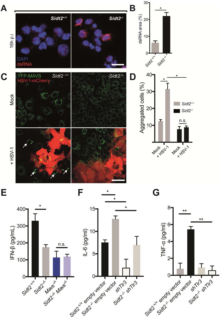
Double-stranded RNA (dsRNA) is a common by-product of viral infections and acts as a potent trigger of antiviral immunity. In the nematode C. elegans, sid-1 encodes a dsRNA transporter that is highly conserved throughout animal evolution, but the physiological role of SID-1 and its orthologs remains unclear. Here, we show that the mammalian SID-1 ortholog, SIDT2, is required to transport internalized extracellular dsRNA from endocytic compartments into the cytoplasm for immune activation. Sidt2-deficient mice exposed to extracellular dsRNA, encephalomyocarditis virus (EMCV), and herpes simplex virus 1 (HSV-1) show impaired production of antiviral cytokines and-in the case of EMCV and HSV-1-reduced survival. Thus, SIDT2 has retained the dsRNA transport activity of its C. elegans ortholog, and this transport is important for antiviral immunity.

Keywords: EMCV; HSV; MAVS; SIDT2; bystander immunity; double-stranded RNA; type I interferon; virus infection.


Conflict of interest statement

The authors declare no competing financial interests.

Figures

Figure 1
Figure 1. SIDT2 localises to late endosomes and interacts with internalised poly(I:C)
(A) MEFs stably expressing SIDT2-mCherry were transfected with markers for (i) early endosomes (EEA1-GFP), (ii) late endosomes (RAB7-GFP) and (iii) lysosomes (LAMP1-YFP), and imaged by confocal microscopy. Co-localization analysis of SIDT2-mCherry with endosomal markers was performed using FIJI software (see main text). Scale bar = 40 μm. (B) DC2.4 cells stably expressing SIDT2-mCherry were transfected with dsDNA-fluorescein or treated with poly(I:C)-fluorescein for 1 h and imaged by confocal microscopy. (C–D) Assessment of SIDT2-mCherry interactions with poly(I:C)-fluorescein and dsDNA-fluorescein in DC2.4 cells via FRET FLIM. ** P < 0.01. Data is plotted as ± SEM and are representative of 3 independent experiments. See also Supplemental videos 1–4.
Figure 2
Figure 2. Loss of SIDT2 impairs innate immunity to EMCV infection
(A) Sidt2+/+ and Sidt2−/− mice (n=6–9) were infected with 50 PFU EMCV i.p. and survival monitored for 8 days. (B–C) Sidt2+/+ and Sidt2−/− mice (n=10–13) were sacrificed at 3 days p.i. and viral load was assessed in the heart and PECs by plaque assay. (D–E) Peritoneal lavage fluid and serum from Sidt2+/+ and Sidt2−/− mice (n=10–13) were collected at day 3 p.i. and IFN-β was measured via ELISA. (F–H) Serum IL-6, RANTES and IL-12 p40 from Sidt2+/+ and Sidt2−/− mice (n=10–13) at 3 days p.i. were measured using Bioplex bead assay. For panels F–H, data are plotted as mean ± SEM. * P < 0.05, *** P < 0.001, n.d = not detected.
Figure 3
Figure 3. Loss of SIDT2 impairs innate immunity to HSV-1 infection
(A) Vero cells were infected with 1 MOI GFP-tagged HSV-1 virus for 24 h, stained with J2 anti-dsRNA antibody (red) and DAPI (blue), and imaged by confocal microscopy. In the absence of HSV-1 infection, no dsRNA was evident (top panel), whereas following HSV-1 challenge dsRNA was readily observed in uninfected cells (bottom panel, arrows). Data are representative of three independent experiments. Scale bar = 20 μm. (B) Sidt2+/+ and Sidt2−/− MEFs were infected with 1 MOI mCherry-tagged HSV-1 virus for the indicated times and analysed by flow cytometry. Data is representative of 3 independent experiments. Error bars represent mean ± SEM. (C) Cell culture supernatant from Sidt2+/+ and Sidt2−/− MEFs infected with 1 MOI mCherry-tagged HSV-1 was collected at 96 h p.i. and IFN-β was measured via ELISA. (D) Sidt2+/+ and Sidt2−/− mice (n=13–15) were infected with 1×10⁷ PFU HSV-1 i.p. and survival monitored for 8 days. (E–F) Serum from Sidt2+/+ and Sidt2−/− mice (n=10–13) was collected at 8h and 16h p.i. respectively and serum IFNβ was measured via ELISA. (G–H) Serum IL-6 and RANTES from Sidt2+/+ and Sidt2−/− mice (n=10–13) at 16 h p.i. were measured using Bioplex bead assay. For panels E–H, data are plotted as mean ± SEM. * P < 0.05, *** P < 0.001, n.s. = not significant. Data represents the pooled results from 3 independent experiments. See also Figure S3.
Figure 4
Figure 4. Loss of SIDT2 impairs endosomal escape of internalised poly(I:C)
BMDCs from Sidt2+/+ and Sidt2−/− mice were treated with (A) poly(I:C)-rhodamine and (B) 32P-labeled 500bp dsRNA for 60 min at either 4°C or 37°C, and internalisation assessed via flow cytometry or radioactivity measurement respectively. Results are representative of at least 2 independent experiments. For panel B, all treatments and measurements were made in triplicate, and data are plotted as mean ± SEM. (C–D) BMDCs from Sidt2+/+ and Sidt2−/− mice were treated with poly(I:C) for either 10 or 60 min, stained with J2 anti-dsRNA antibody (red) and DAPI (blue), and imaged by confocal microscopy. The proportion of each cell occupied by punctate dsRNA staining was quantified. (E–F) Sidt2+/+ and Sidt2−/− BMDCs were treated with poly(I:C) in association with the cationic polymer DEAE-dextran for 60 min. For panels C and E, images are representative of at least three independent experiments. For panels D and F, data are plotted as mean ± SEM and between 30–150 cells were assessed per time point. * P < 0.05. Scale bar = 10 μm. See also Figure S5.
Figure 5
Figure 5. Overexpression of SIDT2 enhances endosomal escape of poly(I:C)
(A) Doxycycline-inducible SIDT2-mCherry DC2.4 cells were treated with poly(I:C)-fluorescein for 1 h in the presence or absence of doxycycline and imaged by confocal microscopy. Scale bar = 10 μm (B) The proportion of each cell occupied by punctate poly(I:C)-fluorescein staining was individually quantified for >60 cells across multiple fields of view, and plotted as mean ± SEM. (C) Internalisation of poly(I:C)-fluorescein by SIDT2-mCherry+ and SIDT2-mCherry cells after 1 h at either 4°C or 37°C was assessed via flow cytometry. Data are representative of 3 independent experiments. ****P < 0.0001. See also Figure S5.
Figure 6
Figure 6. SIDT2 is important for RLR- but not TLR-mediated IFN production in response to extracellular dsRNA
(A) Sidt2+/+ and Sidt2−/− mice were injected i.p with 50 μg poly(I:C) per 25 g body weight (n=7), and serum IFNβ measured at 3 h via ELISA. (B) Sidt2+/+ and Sidt2−/− mice (n=8) were injected i.p. with 300 μg poly(A:U) per 25 g body weight and serum IFNβ measured at 3 h via ELISA. (C) Sidt2+/+ and Sidt2−/− mice (n=8) were injected i.p. with 50 μg poly(I:C) per 25 g body weight and serum IFNλ measured at 3 h via ELISA (n=8). Data are plotted as mean ± SEM. (D) BMDMs from Sidt2+/+ and Sidt2−/− were stimulated with 10 μg/ml poly(I:C) for the indicated times and pIRF3Ser386 and total IRF3 was assessed via immunoblotting. (E) Sidt2+/+ and Sidt2−/− MEFs were transfected with MAVS-YFP, treated with poly(I:C) for 1 h, and MAVS aggregation assessed via confocal microscopy. Scale bar = 40 μm. (F) Individual cells (>150 per condition) were scored for the appearance of MAVS aggregates. Data is representative of 3 independent experiments and error bars are plotted as mean ± SEM. * P < 0.05, n.s. = not significant, n.d = not detected.
Figure 7
Figure 7. SIDT2 is required for RLR-induced MAVS activation in bystander cells during HSV-1 infection
(A) PECs from Sidt2+/+ and Sidt2−/− mice infected with 1×10⁷ PFU at 16 h p.i. were stained with J2 dsRNA antibody (red) and DAPI (blue) and imaged by confocal microscopy. Scale bar = 10 μm. (B) The proportion of each cell occupied by punctate dsRNA staining was quantified using FIJI software. (C) Sidt2+/+ and Sidt2−/− MEFs stably expressing MAVS-YFP were infected with 1 MOI HSV-1-mCherry for 48 h, and imaged via confocal microscopy to assess for MAVS aggregation (arrows) in uninfected, bystander cells. Scale bar = 80 μm. (D) Individual cells were segmented and HSV-1-mCherry infected cells were excluded using FIJI software. Uninfected bystander cells (250 cells per condition) were scored for the appearance of MAVS aggregates. (E) Sidt2+/+ (Cas9 only), Sidt2−/− (Cas9 only), MAVS−/− and Sidt2−/− MAVS−/− MEFs were infected with 1 MOI mCherry-tagged HSV-1, and IFNβ was measured in cell culture supernatant at 96 h p.i. via ELISA. (F) IL-6 and (G) TNF-α were measured in cell culture supernatant from shTLR3 and Sidt2−/− shTLR3 MEFs infected with 1 MOI mCherry-tagged HSV-1 96h p.i. Sidt2+/+ and Sidt2−/− MEFs transduced with retroviral vector lacking shRNA were used as controls. IFN-β, IL-6 and TNF-α in non-infected cells were below the limit of assay detection. Data is representative of results from 2–3 independent experiments and expressed as the mean ± SEM of triplicate wells. * P < 0.05, ** P < 0.01, n.s. = not significant. See also Figure S6.

References

    1. Aubrey BJ, Kelly GL, Kueh AJ, Brennan MS, O’Connor L, Milla L, Wilcox S, Tai L, Strasser A, Herold MJ. An inducible lentiviral guide RNA platform enables the identification of tumor-essential genes and tumor-promoting mutations in vivo. Cell Rep. 2015;10:1422–1432. - PubMed
    1. Barbosa-Morais NL, Irimia M, Pan Q, Xiong HY, Gueroussov S, Lee LJ, Slobodeniuc V, Kutter C, Watt S, Colak R, et al. The evolutionary landscape of alternative splicing in vertebrate species. Science. 2012;338:1587–1593. - PubMed
    1. Barral PM, Sarkar D, Fisher PB, Racaniello VR. RIG-I is cleaved during picornavirus infection. Virology. 2009;391:171–176. - PMC - PubMed
    1. Buschow SI, Lasonder E, Szklarczyk R, Oud MM, de Vries IJ, Figdor CG. Unraveling the human dendritic cell phagosome proteome by organellar enrichment ranking. Journal of proteomics. 2012;75:1547–1562. - PubMed
    1. Chevrier N, Mertins P, Artyomov MN, Shalek AK, Iannacone M, Ciaccio MF, Gat-Viks I, Tonti E, DeGrace MM, Clauser KR, et al. Systematic discovery of TLR signaling components delineates viral-sensing circuits. Cell. 2011;147:853–867. - PMC - PubMed

MeSH terms