. 2017 Sep 19;47(3):524-537.e3.
doi: 10.1016/j.immuni.2017.08.006. Epub 2017 Sep 12.

Glycans Function as Anchors for Antibodies and Help Drive HIV Broadly Neutralizing Antibody Development

Affiliations

Glycans Function as Anchors for Antibodies and Help Drive HIV Broadly Neutralizing Antibody Development

Raiees Andrabi et al. Immunity. .

Erratum in

Abstract

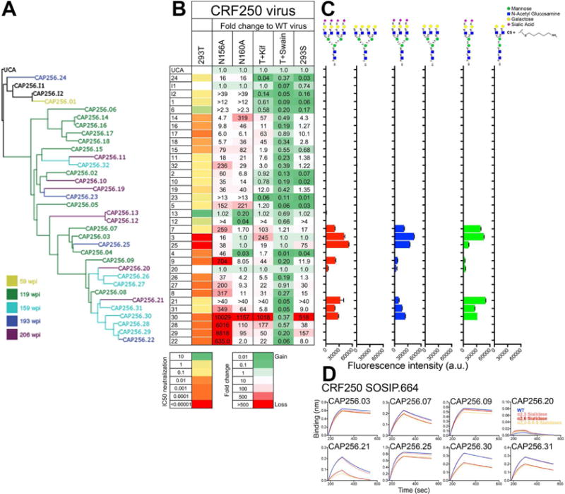
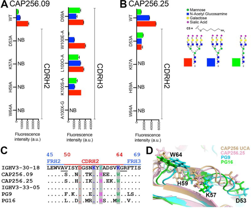
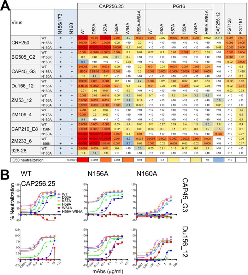
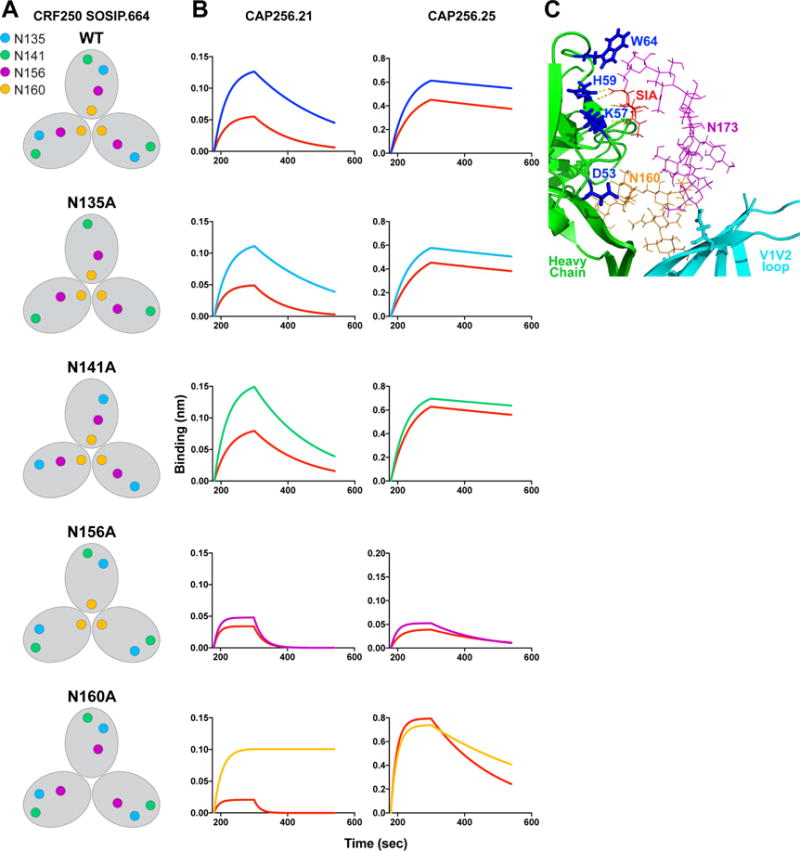
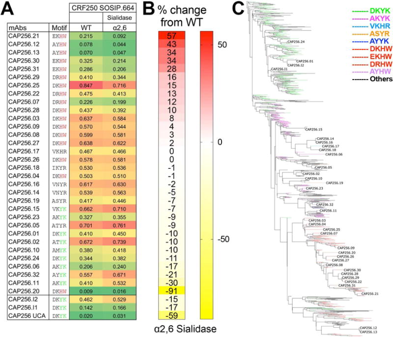
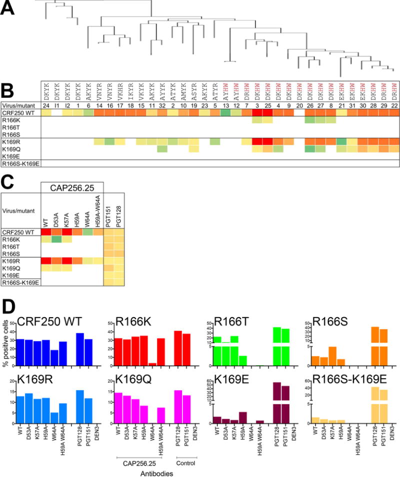
Apex broadly neutralizing HIV antibodies (bnAbs) recognize glycans and protein surface close to the 3-fold axis of the envelope (Env) trimer and are among the most potent and broad Abs described. The evolution of apex bnAbs from one donor (CAP256) has been studied in detail and many Abs at different stages of maturation have been described. Using diverse engineering tools, we investigated the involvement of glycan recognition in the development of the CAP256.VRC26 Ab lineage. We found that sialic acid-bearing glycans were recognized by germline-encoded and somatically mutated residues on the Ab heavy chain. This recognition provided an "anchor" for the Abs as the core protein epitope varies, prevented complete neutralization escape, and eventually led to broadening of the response. These findings illustrate how glycan-specific maturation enables a human Ab to cope with pathogen escape mechanisms and will aid in optimization of immunization strategies to induce V2 apex bnAb responses.

Keywords: B cell affinity maturation; Env glycans; HIV envelope trimer; V2 apex epitope; bnAbs; broadly neutralizing antibodies; glycan engineering; virus escape.


Figures

Figure 1
Figure 1. CAP256 lineage Abs show varying glycan specificity and sialic acid reactivity as measured by neutralization, glycan array and Env trimer binding
A. A phylogenetic tree based on the variable heavy (VH) chain region of CAP256 lineage monoclonal antibodies (CAP256.01-32, I1 and I2). The tree originates from the VH3-30*18 germline gene present in CAP256 UCA. The Abs are colored based on time of sampling (wpi: weeks post infection) B. Heatmap representation of the IC50 neutralization titers of the CAP256 Abs with CRF250 wild-type virus (293T). Fold changes in the IC50 neutralization titers of the CAP256 bnAbs for CRF250 glycan eliminated variants at positions N156 (N156A) and N160 (N160A) and viral glycovariants produced in presence of glycosidase inhibitors (kifunensine (Kif) and swainsonine (Swain)) or in a 293S cell line, which lacks an N-acetyl glucosaminyl transferase enzyme. The mAbs were tested in a TZM-bl cell based infectivity assay (Ab concentration range (IC50 < 0.00001 or >10 μg/ml) and the neutralization IC50 fold changes are represented in heatmap format as indicated in the key. An IC50 fold change value of 1.0 indicates that IC50 neutralization titer is unchanged between the WT virus (293T) and the virus variant. C. Reactivity of the CAP256 Abs to glycans on glycan microarrays. Binding is represented as fluorescence intensity (a.u.). The symbols for each monosaccharide are indicated. D. Octet binding response of the CAP256 bnAbs to CRF250 WT trimer and its three desialylated forms (α2,3 or α2,6 alone or α2,3,6,8,9 cocktail sialidase-treated trimers). The mAbs were immobilized onto an anti-human IgG-Fc sensor followed by dipping these biosensors into the trimer solution and the Ab-trimer interaction is represented as binding curves showing the association (120 seconds; 180–300) and dissociation (240 seconds; 300–540). See also Figure S1 and S2
Figure 2
Figure 2. CDRH2 germline encoded and somatically mutated residues of CAP256.09 and CAP256.25 Abs are important in sialic acid recognition on glycan arrays
A. Reactivity on glycan microarrays of CAP256.09 wild type (WT) antibody and 9 variants. The 9 amino acid substitutions in the CAP256.09 heavy chain variable region include 4 in CDRH2 and 5 in CDRH3. B. Binding of CAP256.25 WT, CDRH2 Ab variants to glycan on the glycan microarray. C. Amino acid sequence alignment of the CDRH2 and parts of the FRH2 and FRH3 region of CAP256.09 and PG9 V2 apex bnAb prototypes with their respective germline VH-genes. Kabat numbering is indicated. The alignment shows that both V2 apex bnAb prototypes retain the germline-encoded residues at CDRH2 positions D53 and K57 and accumulate two common somatic mutations (Y59H and K64W). D. Structural alignment of CAP256 UCA (wheat), CAP256.25 (light pink), PG9 (cyan) and PG16 (green) bnAbs highlighting 4 amino acid positions in the CDRH2 region with side chains as sticks. See also Figure S3
Figure 3
Figure 3. Neutralization of a panel of viruses and glycovariant viruses suggests that CAP256.25 Ab CDRH2 residues interact with sialic acid likely on N156 glycan
A. Neutralization of a panel of viruses including N156 or 173A and N160A variants by CAP256.25 and PG16 Abs and their CDRH2 variants. Neutralization heat maps based on the IC50 titers show the effect of glycan removals on antibody neutralizing activity. PGT128, an N332-V3 Ab and PGT151, a gp120-41 interface Ab, were used as controls. B. Neutralization titration of CAP256.25 WT Ab and its CDRH2 substituted variants against two representative viruses, CAP45_G3 and Du156.12 and their glycan eliminated N156A, N160A variants.
Figure 4
Figure 4. Binding of select CAP256 Abs to CRF250 trimer V2 apex glycan variants shows varying sensitivity to α2,6 sialidase treatment
A. Schematic representation of the V2 apex individual glycan substituted variants on CRF250 SOSIP.664 trimer backbone. B. Octet binding curves (association: 120 s; (180–300) and dissociation: 240 s; (300–540)) of CAP256.21 and CAP256.25 Abs with CRF250 trimer glycovariants. The binding response of CAP256.25 bnAb with the CRF250 N156A variant is fixed to 0.2 nanometer (nm) for clarity C. Ribbon representation of PG16 antibody (green) in complex with isolate ZM109F V1V2 loop (cyan) on a scaffold (modified from (Pancera et al., 2013)). The glycans at N160 (orange; shown as lines) and N173 (magenta; shown as lines) positions and the sialic acid residue (SIA; red) are labeled. The PG16 heavy chain CDRH2 residues (D53, K57, H59 and W64) are highlighted (blue). The residues K57 and H59 of the PG16 heavy chain show hydrogen bonding (yellow dotted line) with a terminal sialic acid residue of N173 glycan. See also Figure S4
Figure 5
Figure 5. Binding of the full range of CAP256 lineage Abs to CRF250 SOSIP.664 shows a gradation of sensitivity to α2,6 sialidase treatment dependent upon key HCDR2 residues
A. Octet binding of CAP256-derived mAbs to PGT145 Ab-affinity purified CRF250 SOSIP.664 trimer (WT) and α2,6 desialylated version. The CAP256 mAbs were captured on an anti-human IgG-Fc sensor and then the CRF250 trimer forms (100nM) flowed over. The binding response (nanometers (nm)) to WT and the α2,6 desialylated trimers is shown as color-coded numerical values and the CAP256 mAbs are arranged with respect to their binding sensitivity to α2,6 desialylated CRF250 trimer. The four amino acids position in the CDRH2 (with respect to germline positions D53, K57, Y59 and K64) are indicated for each Ab. B. Percent (%) change in the octet binding responses are shown as heatmap for the desialylated trimer form and is calculated as (Ab binding response to the α2,6 desialylated CRF250 trimer/binding response to the CRF250 WT trimer). C. Phylogenetic tree showing the development of the CAP256 bnAb lineage from the VH3-30*18 germline gene. The branches in the tree are color-coded and demonstrate the evolution of amino acid residues at the above-mentioned four positions in CDRH2. The germline residues, DKYK and the most evolved residues DKHW in glycan-reactive Abs are indicated in green and red respectively. See also Figure S2C
Figure 6
Figure 6. Sialic acid-binding residues on CAP256.25 bnAb are critical in resisting virus escape via mutations in the basic residue-rich region of V2
A. Phylogenetic tree constructed from VH sequences displaying members of the CAP256 bnAb lineage evolved from the VH3-30*18 germline gene. B. Neutralization heatmap based on the IC50 neutralization titers of CAP256 Ab lineage members with CRF250 WT virus and its 166 and 169 position (HXB2 numbering)-substituted variants that represent escape mutations of CAP256 Abs in natural infection (Bhiman et al., 2015). The 4 CDRH2 amino acid positions, important for glycan recognition, are shown for each Ab member. The lineage members that possess an H and W amino acid residues at positions 59 and 64 respectively are highlighted in red. C. Neutralization heatmap of CAP256.25 Ab and its CDRH2 variants against CRF250 WT virus and positions 166 and 169-substituted variant viruses. D. Binding of CAP256.25 WT Ab and variants to 293T cell-surface expressed CRF250 WT Env and putative escape variant Env trimers. The Abs were used at a final concentration of 10 μg/ml and the extent of binding to the surface trimers is represented as % positive cells as detected by PE-conjugated anti-human-Fc secondary Ab. The V3-N332 high mannose glycan specific Ab PGT128, the gp120–41 trimer cleavage specific Ab PGT151 and a non-HIV Env epitope specific Ab DEN3 were controls for the assay. See also Figure S5 and S6

References

    1. Amin MN, McLellan JS, Huang W, Orwenyo J, Burton DR, Koff WC, Kwong PD, Wang LX. Synthetic glycopeptides reveal the glycan specificity of HIV-neutralizing antibodies. Nature chemical biology. 2013;9:521–526. - PMC - PubMed
    1. Andrabi R, Voss JE, Liang CH, Briney B, McCoy LE, Wu CY, Wong CH, Poignard P, Burton DR. Identification of Common Features in Prototype Broadly Neutralizing Antibodies to HIV Envelope V2 Apex to Facilitate Vaccine Design. Immunity. 2015;43:959–973. - PMC - PubMed
    1. Behrens AJ, Vasiljevic S, Pritchard LK, Harvey DJ, Andev RS, Krumm SA, Struwe WB, Cupo A, Kumar A, Zitzmann N, et al. Composition and Antigenic Effects of Individual Glycan Sites of a Trimeric HIV-1 Envelope Glycoprotein. Cell reports. 2016;14:2695–2706. - PMC - PubMed
    1. Bhiman JN, Anthony C, Doria-Rose NA, Karimanzira O, Schramm CA, Khoza T, Kitchin D, Botha G, Gorman J, Garrett NJ, et al. Viral variants that initiate and drive maturation of V1V2-directed HIV-1 broadly neutralizing antibodies. Nature medicine. 2015;21:1332–1336. - PMC - PubMed
    1. Bonsignori M, Hwang KK, Chen X, Tsao CY, Morris L, Gray E, Marshall DJ, Crump JA, Kapiga SH, Sam NE, et al. Analysis of a clonal lineage of HIV-1 envelope V2/V3 conformational epitope-specific broadly neutralizing antibodies and their inferred unmutated common ancestors. Journal of virology. 2011;85:9998–10009. - PMC - PubMed

Publication types

MeSH terms