

Architectural delineation and molecular identification of extracellular matrix in ascidian embryos and larvae
- PMID: 28916708
- PMCID: PMC5612238
- DOI: 10.1242/bio.026336
Architectural delineation and molecular identification of extracellular matrix in ascidian embryos and larvae
Abstract
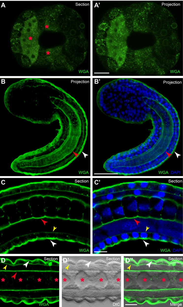
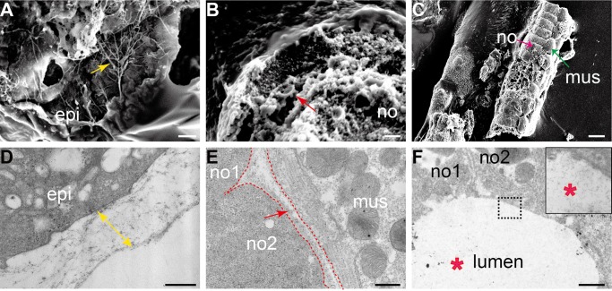
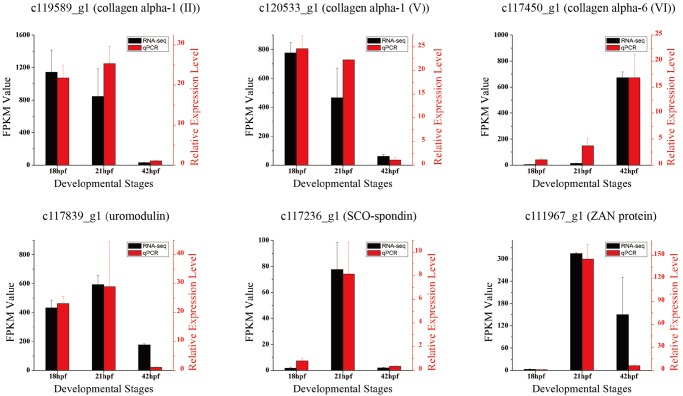
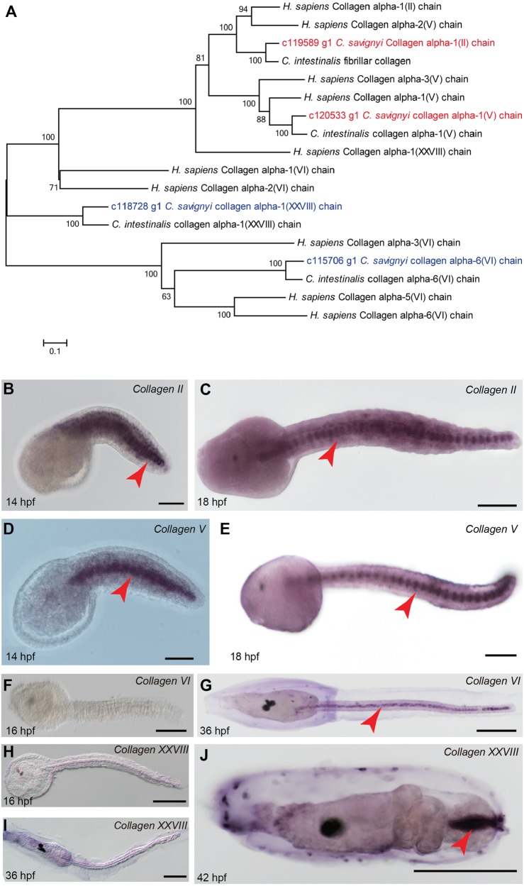
The extracellular matrix (ECM) not only provides essential physical scaffolding for cellular constituents but also initiates crucial biochemical and biomechanical cues that are required for tissue morphogenesis. In this study, we utilized wheat germ agglutinin (WGA) staining to characterize the ECM architecture in ascidian embryos and larvae. The results showed three distinct populations of ECM presenting in Ciona embryogenesis: the outer layer localized at the surface of embryo, an inner layer of notochord sheath and the apical ECM secreted by the notochord. To further elucidate the precise structure of Ciona embryonic ECM, we employed scanning and transmission electron microscopy, and found that the outer membrane was relatively thick with short fibres, whereas the ECM layer in notochord sheath was not as thick as the outer membrane but more regular arranged; the lumen between notochord cells was hydrostatic and sticky. Then, we used the RNA sequencing data from the embryos and larvae of Ciona savignyi to identify ECM genes and acquire their expression patterns. We identified 115 unigenes as 67 ECM genes, and 77 unigenes showed dynamic expression changes between different stages. Our results reveal the architecture, molecular composition and dynamic expression profile of ECM in ascidian embryogenesis, and may increase understanding of the function of the ECM in chordate development.
Keywords: Ciona; Collagen; Extracellular matrix; RNA sequencing.
© 2017. Published by The Company of Biologists Ltd.
Conflict of interest statement
Competing interestsThe authors declare no competing or financial interests.
Figures
Similar articles
-
Dynamics of Chromatin Opening across Larval Development in the Urochordate Ascidian Ciona savignyi.Int J Mol Sci. 2024 Feb 28;25(5):2793. doi: 10.3390/ijms25052793. Int J Mol Sci. 2024. PMID: 38474039 Free PMC article.
-
Identification and expression analysis of long noncoding RNAs in embryogenesis and larval metamorphosis of Ciona savignyi.Mar Genomics. 2018 Jul;40:64-72. doi: 10.1016/j.margen.2018.05.001. Epub 2018 May 10. Mar Genomics. 2018. PMID: 29754835
-
Role of the ECM in notochord formation, function and disease.J Cell Sci. 2017 Oct 1;130(19):3203-3211. doi: 10.1242/jcs.175950. Epub 2017 Sep 7. J Cell Sci. 2017. PMID: 28883093 Review.
-
Tube formation by complex cellular processes in Ciona intestinalis notochord.Dev Biol. 2009 Jun 15;330(2):237-49. doi: 10.1016/j.ydbio.2009.03.015. Epub 2009 Mar 24. Dev Biol. 2009. PMID: 19324030 Free PMC article.
-
Ascidian notochord elongation.Dev Biol. 2019 Apr 15;448(2):147-153. doi: 10.1016/j.ydbio.2018.11.009. Epub 2018 Nov 17. Dev Biol. 2019. PMID: 30458170 Review.
Cited by
-
DYRK1-mediated phosphorylation of endocytic components is required for extracellular lumen expansion in ascidian notochord.Biol Res. 2023 Mar 11;56(1):10. doi: 10.1186/s40659-023-00422-9. Biol Res. 2023. PMID: 36899423 Free PMC article.
-
Biomimetic Layer-by-Layer Self-Assembly of Nanofilms, Nanocoatings, and 3D Scaffolds for Tissue Engineering.Int J Mol Sci. 2018 Jun 1;19(6):1641. doi: 10.3390/ijms19061641. Int J Mol Sci. 2018. PMID: 29865178 Free PMC article. Review.
-
Polarity Establishment and Maintenance in Ascidian Notochord.Front Cell Dev Biol. 2020 Oct 30;8:597446. doi: 10.3389/fcell.2020.597446. eCollection 2020. Front Cell Dev Biol. 2020. PMID: 33195278 Free PMC article. Review.
-
Integrated multi-omics identify key signalling pathways for notochord lumenogenesis in ascidian Ciona savignyi.Open Biol. 2025 Apr;15(4):240402. doi: 10.1098/rsob.240402. Epub 2025 Apr 9. Open Biol. 2025. PMID: 40199341 Free PMC article.
-
Single-cell Transcriptomic Studies Unveil Potential Nodes of the Notochord Gene Regulatory Network.Integr Comp Biol. 2024 Nov 21;64(5):1194-1213. doi: 10.1093/icb/icae084. Integr Comp Biol. 2024. PMID: 38914463 Free PMC article. Review.
References
LinkOut - more resources
Full Text Sources
Other Literature Sources
