Identification of new channels by systematic analysis of the mitochondrial outer membrane
- PMID: 28916712
- PMCID: PMC5674900
- DOI: 10.1083/jcb.201706043
Identification of new channels by systematic analysis of the mitochondrial outer membrane
Abstract
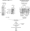
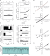
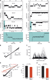
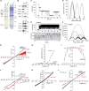
The mitochondrial outer membrane is essential for communication between mitochondria and the rest of the cell and facilitates the transport of metabolites, ions, and proteins. All mitochondrial outer membrane channels known to date are β-barrel membrane proteins, including the abundant voltage-dependent anion channel and the cation-preferring protein-conducting channels Tom40, Sam50, and Mdm10. We analyzed outer membrane fractions of yeast mitochondria and identified four new channel activities: two anion-preferring channels and two cation-preferring channels. We characterized the cation-preferring channels at the molecular level. The mitochondrial import component Mim1 forms a channel that is predicted to have an α-helical structure for protein import. The short-chain dehydrogenase-related protein Ayr1 forms an NADPH-regulated channel. We conclude that the mitochondrial outer membrane contains a considerably larger variety of channel-forming proteins than assumed thus far. These findings challenge the traditional view of the outer membrane as an unspecific molecular sieve and indicate a higher degree of selectivity and regulation of metabolite fluxes at the mitochondrial boundary.
© 2017 Krüger et al.
Figures


Similar articles
-
Mdm10 is an ancient eukaryotic porin co-occurring with the ERMES complex.Biochim Biophys Acta. 2013 Dec;1833(12):3314-3325. doi: 10.1016/j.bbamcr.2013.10.006. Epub 2013 Oct 14. Biochim Biophys Acta. 2013. PMID: 24135058
-
Biogenesis of the mitochondrial TOM complex: Mim1 promotes insertion and assembly of signal-anchored receptors.J Biol Chem. 2008 Jan 4;283(1):120-127. doi: 10.1074/jbc.M706997200. Epub 2007 Nov 1. J Biol Chem. 2008. PMID: 17974559
-
Protein translocase of the outer mitochondrial membrane: role of import receptors in the structural organization of the TOM complex.J Mol Biol. 2002 Feb 22;316(3):657-66. doi: 10.1006/jmbi.2001.5365. J Mol Biol. 2002. PMID: 11866524
-
Mitochondrial Outer Membrane Channels: Emerging Diversity in Transport Processes.Bioessays. 2018 Jul;40(7):e1800013. doi: 10.1002/bies.201800013. Epub 2018 Apr 30. Bioessays. 2018. PMID: 29709074 Review.
-
Structure and evolution of mitochondrial outer membrane proteins of beta-barrel topology.Biochim Biophys Acta. 2010 Jun-Jul;1797(6-7):1292-9. doi: 10.1016/j.bbabio.2010.04.019. Epub 2010 May 5. Biochim Biophys Acta. 2010. PMID: 20450883 Review.
Cited by
-
Post-translational modifications and protein quality control of mitochondrial channels and transporters.Front Cell Dev Biol. 2023 Aug 3;11:1196466. doi: 10.3389/fcell.2023.1196466. eCollection 2023. Front Cell Dev Biol. 2023. PMID: 37601094 Free PMC article. Review.
-
Role of Mitochondrial Dysfunctions in Neurodegenerative Disorders: Advances in Mitochondrial Biology.Mol Neurobiol. 2025 Jun;62(6):6827-6855. doi: 10.1007/s12035-024-04469-x. Epub 2024 Sep 13. Mol Neurobiol. 2025. PMID: 39269547 Review.
-
Mitochondrial protein translocation machinery: From TOM structural biogenesis to functional regulation.J Biol Chem. 2022 May;298(5):101870. doi: 10.1016/j.jbc.2022.101870. Epub 2022 Mar 26. J Biol Chem. 2022. PMID: 35346689 Free PMC article. Review.
-
The Role of Oxidative Stress as a Mechanism in the Pathogenesis of Acute Heart Failure in Acute Kidney Injury.Diagnostics (Basel). 2024 Sep 23;14(18):2094. doi: 10.3390/diagnostics14182094. Diagnostics (Basel). 2024. PMID: 39335773 Free PMC article. Review.
-
Role of the Mitochondrial Protein Import Machinery and Protein Processing in Heart Disease.Front Cardiovasc Med. 2021 Sep 28;8:749756. doi: 10.3389/fcvm.2021.749756. eCollection 2021. Front Cardiovasc Med. 2021. PMID: 34651031 Free PMC article. Review.
References
MeSH terms
Substances
Grants and funding
LinkOut - more resources
Full Text Sources
Other Literature Sources
Molecular Biology Databases

