. 2017 Nov 3;292(44):18240-18255.
doi: 10.1074/jbc.M117.813527. Epub 2017 Sep 15.

Phosphoproteomics reveals that glycogen synthase kinase-3 phosphorylates multiple splicing factors and is associated with alternative splicing

Affiliations

Phosphoproteomics reveals that glycogen synthase kinase-3 phosphorylates multiple splicing factors and is associated with alternative splicing

Mansi Y Shinde et al. J Biol Chem. .

Abstract

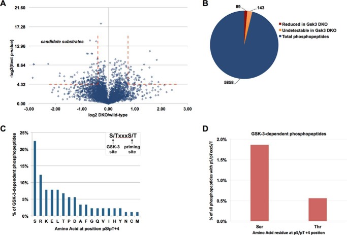
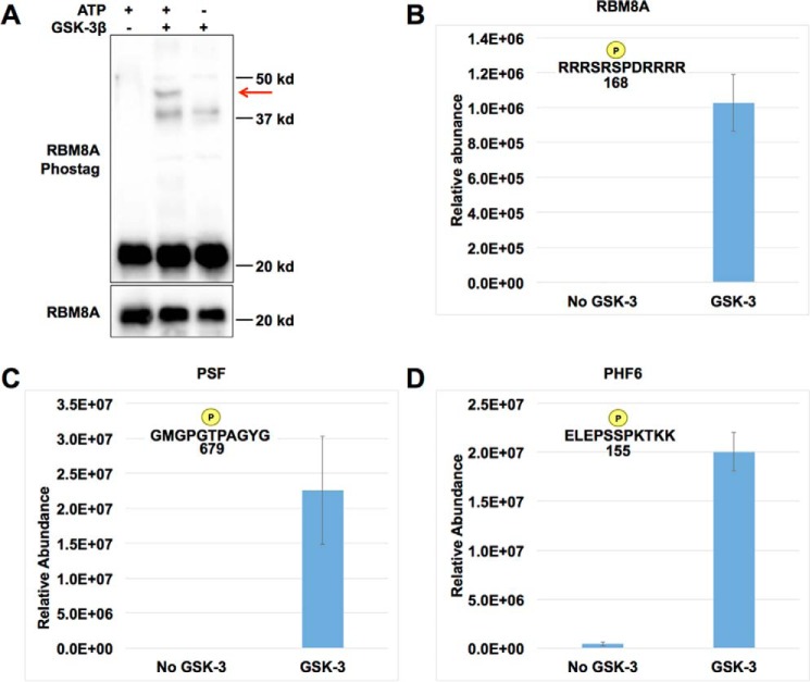
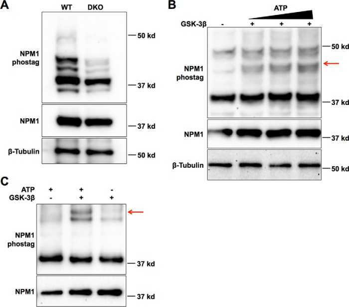
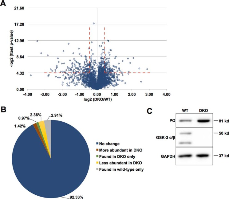
Glycogen synthase kinase-3 (GSK-3) is a constitutively active, ubiquitously expressed protein kinase that regulates multiple signaling pathways. In vitro kinase assays and genetic and pharmacological manipulations of GSK-3 have identified more than 100 putative GSK-3 substrates in diverse cell types. Many more have been predicted on the basis of a recurrent GSK-3 consensus motif ((pS/pT)XXX(S/T)), but this prediction has not been tested by analyzing the GSK-3 phosphoproteome. Using stable isotope labeling of amino acids in culture (SILAC) and MS techniques to analyze the repertoire of GSK-3-dependent phosphorylation in mouse embryonic stem cells (ESCs), we found that ∼2.4% of (pS/pT)XXX(S/T) sites are phosphorylated in a GSK-3-dependent manner. A comparison of WT and Gsk3a;Gsk3b knock-out (Gsk3 DKO) ESCs revealed prominent GSK-3-dependent phosphorylation of multiple splicing factors and regulators of RNA biosynthesis as well as proteins that regulate transcription, translation, and cell division. Gsk3 DKO reduced phosphorylation of the splicing factors RBM8A, SRSF9, and PSF as well as the nucleolar proteins NPM1 and PHF6, and recombinant GSK-3β phosphorylated these proteins in vitro RNA-Seq of WT and Gsk3 DKO ESCs identified ∼190 genes that are alternatively spliced in a GSK-3-dependent manner, supporting a broad role for GSK-3 in regulating alternative splicing. The MS data also identified posttranscriptional regulation of protein abundance by GSK-3, with ∼47 proteins (1.4%) whose levels increased and ∼78 (2.4%) whose levels decreased in the absence of GSK-3. This study provides the first unbiased analysis of the GSK-3 phosphoproteome and strong evidence that GSK-3 broadly regulates alternative splicing.

Keywords: SILAC; Wnt signaling; alternative splicing; embryonic stem cell; glycogen synthase kinase 3 (GSK-3); leukemia; lithium; phosphoproteomics; spliceosome.


Conflict of interest statement

The authors declare that they have no conflicts of interest with the contents of this article

Figures

Figure 1.
Figure 1.
Global phosphoproteome analysis using SILAC. A, experimental work flow. Cells were cultured for four passages in medium supplemented with either heavy (Arg 10/Lys 8) or light (Arg 0/Lys 0) amino acids to ensure complete incorporation. Whole-cell lysates were digested with trypsin, and phosphorylated peptides were enriched using titanium oxide columns. Flow-through proteins were used to identify the proteome. B, flow cytometry for the ESC pluripotency markers Oct4 and Nanog indicates homogeneity of the wild-type and DKO cell populations. C, Western blotting to assess Gsk3 knock-out (middle) and Wnt signaling activation by β-catenin protein accumulation (top). GAPDH is a loading control. D, overview of phosphorylation sites identified in the screen.
Figure 2.
Figure 2.
Gsk3 null ESC phosphoproteome identifies candidate substrates of GSK-3. A, volcano plot showing log2(-fold change) of each phosphopeptide (DKO/wild type) versus −log2(p value). Broken red lines indicate peptides with an absolute change of 1.5-fold or greater, p value ≤ 0.05. B, distribution of phosphopeptides that were significantly reduced in Gsk3 DKO (red), phosphopeptides present in wild type but undetectable in DKO (yellow), and phosphopeptides that did not change significantly (blue). C, frequency of each amino acid at the +4-position relative to the GSK-3–dependent phosphorylation site (inset shows reported GSK-3 consensus sequence with serine or threonine at +4-position (potential priming sites)). D, frequency of GSK-3–dependent phosphopeptides with serine (1.9%) or threonine (0.6%) at the +4-position as a percentage of all 1075 identified phosphopeptides with serine or threonine at the +4-position.
Figure 3.
Figure 3.
Proteins with a broad range of functions are phosphorylated in a GSK-3–dependent manner in mouse ESCs. A, gene ontology analysis of proteins with significantly reduced phosphorylation in Gsk3 DKO cells using DAVID. B, STRING analysis of proteins enriched in the GO analysis generates three networks of proteins with related functions. Darker hue indicates greater reduction in phosphorylation in DKO cells. The red asterisk indicates phosphorylation sites with GSK-3 consensus motif. Phosphorylation of proteins in this analysis was reduced >1.5-fold with p value ≤ 0.05.
Figure 4.
Figure 4.
GSK-3 directly phosphorylates RBM8A, PSF, and PHF6. A, in vitro GSK-3–dependent phosphorylation of recombinant RBM8A protein (red arrow) resolved by Phos-tag polyacrylamide electrophoresis and Western blotting. B–D, mass spectrometry demonstrates GSK-3 phosphorylation of RBM8A at serine 168 (B), PSF at threonine 679 (C), and PHF6 at serine 155 (D). Error bars, S.D.
Figure 5.
Figure 5.
GSK-3 directly phosphorylates NPM1. A, multiple phosphorylated forms of NPM1 were detected by Western blotting of wild-type and Gsk3 DKO cell lysates resolved by Phos-tag gel (top). NPM1 migrates as a single species in standard electrophoresis (middle). Bottom, β-tubulin loading control. B, Gsk3 DKO cell lysates were added to an in vitro protein kinase reaction with recombinant GSK-3 with or without ATP for 1 h. The reaction was then resolved by Phos-tag gel, and NPM1 was visualized by Western blotting. A slower-migrating species detected only in the presence of ATP, representing a GSK-3–phosphorylated form of NPM1, is indicated by the red arrow. C, GSK-3 phosphorylation of recombinant NPM1 in vitro resolved by Phos-tag (top) and standard electrophoresis (bottom).
Figure 6.
Figure 6.
Analysis of gene expression changes in wild-type and Gsk3 DKO mESCs. A, work flow of alternative splicing (AS) and differential expression (DE) analysis from RNA-Seq. B, total number of genes differing in alternative splicing or differential expression between wild-type and DKO cells. The six genes observed in both populations are indicated below the Venn diagram. C, categories of alternative splicing for the 194 changing splicing events detected by MAJIQ. Categories are defined as indicated by the schematics, followed by number of events in that category. Events not falling in any single category are defined as “Complex.”
Figure 7.
Figure 7.
Protein abundance changes in DKO compared with wild type. A, volcano plot of log2(-fold change in protein abundance) in DKO/wild-type versus −log2(p value) demonstrates multiple proteins that are both increased and decreased in abundance in a Gsk3 dependent manner. B, percentage of proteins that are increased (red) or decreased (yellow) 1.5-fold (p < 0.05) in abundance in Gsk3 DKO cells without a statistically significant change in RNA abundance. The percentage of proteins detected only in DKO cells (green) or only in Gsk3 wild-type ESCs (gray) without a statistically significant change in RNA abundance is also shown. C, PREP/PO total protein levels are increased in DKO compared with wild type without a significant change in RNA abundance.

References

    1. Doble B. W., and Woodgett J. R. (2003) GSK-3: tricks of the trade for a multi-tasking kinase. J. Cell Sci. 116, 1175–1186 - PMC - PubMed
    1. Picton C., Woodgett J., Hemmings B., and Cohen P. (1982) Multisite phosphorylation of glycogen synthase from rabbit skeletal muscle: phosphorylation of site 5 by glycogen synthase kinase-5 (casein kinase-II) is a prerequisite for phosphorylation of sites 3 by glycogen synthase kinase-3. FEBS Lett. 150, 191–196 - PubMed
    1. Hoeflich K. P., Luo J., Rubie E. A., Tsao M. S., Jin O., and Woodgett J. R. (2000) Requirement for glycogen synthase kinase-3β in cell survival and NF- κB activation. Nature 406, 86–90 - PubMed
    1. Liu K. J., Arron J. R., Stankunas K., Crabtree G. R., and Longaker M. T. (2007) Chemical rescue of cleft palate and midline defects in conditional GSK-3β mice. Nature 446, 79–82 - PubMed
    1. Barrell W. B., Szabo-Rogers H. L., and Liu K. J. (2012) Novel reporter alleles of GSK-3α and GSK-3β. PLoS One 7, e50422. - PMC - PubMed

MeSH terms