. 2017 Oct 20;292(42):17324-17336.
doi: 10.1074/jbc.M117.788331. Epub 2017 Sep 15.

A novel mechanism for Ca2+/calmodulin-dependent protein kinase II targeting to L-type Ca2+ channels that initiates long-range signaling to the nucleus

Affiliations

A novel mechanism for Ca2+/calmodulin-dependent protein kinase II targeting to L-type Ca2+ channels that initiates long-range signaling to the nucleus

Xiaohan Wang et al. J Biol Chem. .

Abstract

Neuronal excitation can induce new mRNA transcription, a phenomenon called excitation-transcription (E-T) coupling. Among several pathways implicated in E-T coupling, activation of voltage-gated L-type Ca2+ channels (LTCCs) in the plasma membrane can initiate a signaling pathway that ultimately increases nuclear CREB phosphorylation and, in most cases, expression of immediate early genes. Initiation of this long-range pathway has been shown to require recruitment of Ca2+-sensitive enzymes to a nanodomain in the immediate vicinity of the LTCC by an unknown mechanism. Here, we show that activated Ca2+/calmodulin-dependent protein kinase II (CaMKII) strongly interacts with a novel binding motif in the N-terminal domain of CaV1 LTCC α1 subunits that is not conserved in CaV2 or CaV3 voltage-gated Ca2+ channel subunits. Mutations in the CaV1.3 α1 subunit N-terminal domain or in the CaMKII catalytic domain that largely prevent the in vitro interaction also disrupt CaMKII association with intact LTCC complexes isolated by immunoprecipitation. Furthermore, these same mutations interfere with E-T coupling in cultured hippocampal neurons. Taken together, our findings define a novel molecular interaction with the neuronal LTCC that is required for the initiation of a long-range signal to the nucleus that is critical for learning and memory.

Keywords: Ca2+/calmodulin-dependent protein kinase II (CaMKII); cAMP-response element-binding protein (CREB); calcium channel; neuron; protein-protein interaction.


Conflict of interest statement

The authors declare that they have no conflicts of interest with the contents of this article

Figures

Figure 1.
Figure 1.
Activated CaMKII specifically binds to the LTCC NTD. A, domain structure of LTCCs and GST fusion proteins used here. Rectangular boxes in the intracellular domains indicate approximate positions of previously reported calmodulin (NSCaTE (24)) (purple box)-binding and CaMKII (20) (white box)-binding domains in the NTD, the α subunit interaction domain (AID, for β subunit interaction) in the I/II linker (50) (blue box), and overlapping calmodulin- and CaMKII-binding sites in the CTD (17) (green box). B, glutathione-agarose co-sedimentation assays show that there is no reliably detectable interaction of inactive (non-autophosphorylated) conformations of CaMKIIα with any of the CaV1.3 intracellular domains but that activated (pre-autophosphorylated) CaMKIIα specifically binds to the NTD. C, activation of CaMKIIα by binding of Ca2+/calmodulin and Mg-ADP is sufficient for interaction with the CaV1.3 NTD. The immunoblots shown are representative of three independent experiments.
Figure 2.
Figure 2.
CaMKII specifically binds to LTCC NTDs. A, alignment of membrane-proximal regions of the NTDs from all human VGCCs. Ca2+ channel NTDs are more conserved in membrane-proximal regions but become more divergent in the distal regions. B, representative glutathione-agarose co-sedimentation assay comparing the binding of activated CaMKII to GST-NTDs from CaV1, CaV2, and CaV3 Ca2+ channels. C, quantitation of 2–3 independent experiments similar to those shown in B. The CaV1.3 NTD shows the strongest binding to CaMKIIα, followed by CaV1.2, whereas interactions with CaV2.2 and CaV3.2 are barely detected. All values were normalized to CaV1.3 NTD pulldown. Error bars, S.E.
Figure 3.
Figure 3.
Characterization of the CaV1.3 NTD CaMKII-binding domain. A, truncations used to map the CaMKII interaction site in the CaV1.3 NTD. Purple and white rectangles indicate approximate positions of previously defined NSCaTE calmodulin-binding and CaMKII-binding domains, respectively (see legend to Fig. 1 and “Results”). B, glutathione-agarose co-sedimentation assay comparing binding of activated CaMKIIα to the full-length CaV1.3 NTD, the membrane-distal part (NT-A), and the membrane-proximal part (NT-B). C, analysis of further NTD truncations reveals that the NT-B1 region (residues 69–93) is sufficient for binding of activated CaMKIIα. D, mutation of amino acids 83RKR85 to AAA within the full-length CaV1.3 NTD blocks CaV1.3–CaMKIIα interaction. These immunoblots are representative of at least three independent replicates.
Figure 4.
Figure 4.
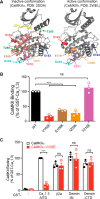
Identification of a CaMKII mutation that specifically disrupts binding to the CaV1.3 NTD. A, CaMKII structures. Left, a single CaMKIIα subunit in an inactive (autoinhibited) conformation with an inhibitor (Bosutinib, yellow) bound in the nucleotide binding site (PDB entry 3SOA (51)). Right, a single CaMKIIδ subunit in an activated conformation (displaced regulatory domain) with a bound inhibitor (SU6656, yellow) (PDB entry 2WEL (52)). The catalytic and regulatory domains are shown in gray and pink, respectively. For clarity of presentation, C-terminal holoenzyme association domains are not shown, and the displaced regulatory domain with bound Ca2+/calmodulin is not shown in PDB entry 2WEL. Thr286 and Thr305 (green) are two regulatory autophosphorylation sites. Mutation of Ile205/206 (orange) to Lys disrupts CaMKII interaction with GluN2B and densin-IN (22, 26), whereas mutation of Asp238/239 (cyan) to Arg disrupts GluN2B binding but spares interactions with densin-IN (22). A naturally occurring de novo Glu183 (purple) to Val mutation in CaMKIIα is linked to autism spectrum disorder and disrupts CaMKII interaction with multiple CaMKAPs (48, 53). B, a 96-well glutathione plate assay to screen activated mApple-tagged CaMKIIα mutants for interactions with GST-tagged CaV1.3 NTD. C, binding of activated mApple-tagged WT and V102E-CaMKIIα to multiple GST-CaMKAP proteins in the 96-well plate assay. A V102E mutation selectively disrupts CaMKIIα binding to the CaV1.3 NTD; Val102/103 is highlighted in red in A. Data from three independent experiments were analyzed by one-way ANOVA (for B) and two-way ANOVA followed by Sidak's multiple-comparison test (for C), respectively. ***, p < 0.001; ns, not significant (p > 0.05). Error bars, S.E.
Figure 5.
Figure 5.
The NTD is important for CaMKII association with LTCC complexes. A, preactivated CaMKIIα robustly interacts with the minimal CaMKII-binding sites from the CaV1.3 NTD and the β2 auxiliary subunit, but not with a previously reported minimal CaMKII-binding site in the CaV1.2 CTD that is identical in CaV1.3. B, a schematic diagram showing the structure of chimeric CaVx.x NTD-CaV1.3 channels in which the CaV1.3 NTD was substituted by NTDs from CaV2.2 or CaV3.2. C, equal aliquots of lysates from cells expressing CaMKIIα with WT or NTD chimeric HA-tagged CaV1.3s were immunoprecipitated using anti-HA antibodies without (EDTA) or with the addition of excess Ca2+/calmodulin/Mg2+-ATP. C2 plots levels of immunoprecipitated HA-CaV1.3 proteins (black) and CaMKII (purple) in the presence of Ca2+/calmodulin/Mg2+/ATP normalized to levels isolated in the presence of EDTA in each experiment. C3 compares levels of immunoprecipitated CaMKIIα normalized to immunoprecipitated HA proteins in the presence of EDTA and Ca2+/calmodulin/Mg2+-ATP. D, similar analysis of the co-immunoprecipitation of WT or V102E-CaMKIIα with WT, Δ69–93, or RKR-AAA HA-CaV1.3 in the presence of EDTA or Ca2+/calmodulin/Mg2+/ATP. Levels of immunoprecipitated HA-CaV1.3 and CaMKIIα are compared in D2, and normalized CaMKIIα/HA-CaV1.3 ratios are shown in D3. Data are from 3–4 independent experiments and analyzed by two-way ANOVA followed by Sidak's multiple-comparison test. *, p < 0.05; ***, p < 0.001; ns, not significant (p > 0.05). Error bars, S.E.
Figure 6.
Figure 6.
Deletion of residues 69–93 from the CaV1.3 NTD does not affect Ca2+ influx via CaV1.3 LTCCs. A, representative Ca2+ currents elicited by step depolarizations (50 ms) to various voltages for CaV1.3-WT (left) and CaV1.3-Δ69–93 LTCCs. Scale bars, 10 ms (horizontal) and 50 pA (vertical), respectively. B, no significant difference in current-voltage (I-V) relationships for CaV1.3-WT and CaV1.3-Δ69–93 LTCCs (p > 0.05, two-way ANOVA followed by Sidak's multiple-comparison test). C, no significant difference in peak current densities of CaV1.3-WT and CaV1.3-Δ69–93 LTCCs. D, no significant difference in fast inactivation of CaV1.3-WT and CaV1.3-Δ69–93 LTCCs, based on the fraction of residual current measured 30 ms after depolarization from −70 to 0 mV. Data were collected from five independent transfections, n = 13 for WT and n = 15 for Δ69–93. Data were analyzed by two-tailed unpaired Student's t test; ns, not significant (p > 0.05). Error bars, S.E.
Figure 7.
Figure 7.
Role of the LTCC NTD in high K+-induced CREB Ser133 phosphorylation. A, Ca2+ imaging showing that nimodipine largely prevents the high K+-induced increase of somatic Ca2+. Neurons were incubated with Tyrode's solution containing 5 mm KCl (5K) for 30 s and switched to Tyrode's solution containing 40 mm KCl for 2 min in the absence (40K control, n = 193) or presence (40K + NIM, n = 215) of 10 μm nimodipine. The black arrow indicates the buffer switch, and data were plotted as mean ± S.E. A total of five dishes from two independent cultures were analyzed per group. B, depolarization of cultured hippocampal neurons induced LTCC-dependent Ser133 phosphorylation of CREB. In the columns from left to right, neurons were incubated with 5K Tyrode's solution and then switched to 5K, 40K control, or 40K + NIM Tyrode's solution, respectively, for 90 s. Neurons were then fixed and stained for DAPI, CREB Ser133 phosphorylation (pCREB), and CaMKIIα. Incubation with 40 mm KCl induced an increase of pCREB that was blocked by the LTCC antagonist nimodipine. C, deletion of the CaMKII-binding domain in the CaV1.3 NTD disrupts nuclear signaling. Expression of a nimodipine-resistant CaV1.3-T1033Y mutant rescues the nimodipine blockade of pCREB induction by 40 mm KCl. However, deletion of the CaMKII-binding domain (Δ69–93) prevents the rescue of pCREB signaling by CaV1.3-T1033Y. Each data point represents analysis of a single cell collected from 3–4 independent neuronal cultures/transfections. Pooled data were analyzed by one-way ANOVA followed by Tukey's multiple-comparison test. ***, p < 0.001; ns, not significant (p > 0.05). All confocal images show a 40 × 40-μm area. Error bars, S.E.
Figure 8.
Figure 8.
CaMKII-binding to the CaV1.3 NTD is required for high K+-induced CREB Ser133 phosphorylation. A, CaMKIIα/β knockdown or re-expression/rescue has no effect on high K+-induced increases of somatic Ca2+. Cultured hippocampal neurons were transfected with mApple only (n = 17), mApple with CaMKIIα/β shRNA (n = 23), mApple/CaMKIIα/β shRNA with shRNA-resistant CaMKIIαR-WT (n = 26), or CaMKIIαR-V102E cDNA (n = 17), respectively. Transfected neurons were then monitored for Ca2+ influx in response to 40 mm K+-induced depolarization (black arrow; see Fig. 7A). B, the CaMKIIα-V102E mutant does not support nuclear signaling. Expression of CaMKIIα/CaMKIIβ shRNAs significantly reduces nuclear CREB phosphorylation following 40 mm KCl treatment. This reduction in CREB phosphorylation is rescued by co-expression of shRNA-resistant CaMKIIαR-WT, but not CaMKIIαR-K42R (kinase-dead) or CaMKIIαR-V102E (deficient in CaV1.3 NTD binding). Each data point represents analysis of a single cell collected from 3–4 independent neuronal cultures/transfections. Pooled data were analyzed by one-way ANOVA followed by Tukey's multiple-comparison test. ***, p < 0.001; ns, not significant (p > 0.05). All confocal images show a 40 × 40-μm area. Error bars, S.E.

Similar articles

Cited by

References

    1. Hagenston A. M., and Bading H. (2011) Calcium signaling in synapse-to-nucleus communication. Cold Spring Harb. Perspect. Biol. 3, a004564. - PMC - PubMed
    1. Deisseroth K., Mermelstein P. G., Xia H., and Tsien R. W. (2003) Signaling from synapse to nucleus: the logic behind the mechanisms. Curr. Opin. Neurobiol. 13, 354–365 - PubMed
    1. Bading H. (2013) Nuclear calcium signalling in the regulation of brain function. Nat. Rev. Neurosci. 14, 593–608 - PubMed
    1. Flavell S. W., and Greenberg M. E. (2008) Signaling mechanisms linking neuronal activity to gene expression and plasticity of the nervous system. Annu. Rev. Neurosci. 31, 563–590 - PMC - PubMed
    1. Alberini C. M. (2009) Transcription factors in long-term memory and synaptic plasticity. Physiol. Rev. 89, 121–145 - PMC - PubMed

Publication types

Substances

LinkOut - more resources