

Muscle pathology from stochastic low level DUX4 expression in an FSHD mouse model
- PMID: 28916757
- PMCID: PMC5601940
- DOI: 10.1038/s41467-017-00730-1
Muscle pathology from stochastic low level DUX4 expression in an FSHD mouse model
Erratum in
-
Author Correction: Muscle pathology from stochastic low level DUX4 expression in an FSHD mouse model.Nat Commun. 2018 Feb 22;9(1):856. doi: 10.1038/s41467-018-03449-9. Nat Commun. 2018. PMID: 29472544 Free PMC article.
Abstract
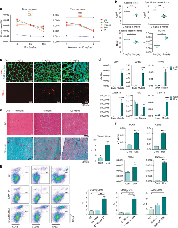
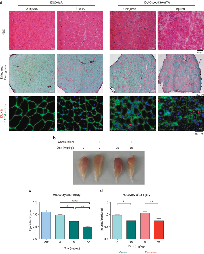
Facioscapulohumeral muscular dystrophy is a slowly progressive but devastating myopathy caused by loss of repression of the transcription factor DUX4; however, DUX4 expression is very low, and protein has not been detected directly in patient biopsies. Efforts to model DUX4 myopathy in mice have foundered either in being too severe, or in lacking muscle phenotypes. Here we show that the endogenous facioscapulohumeral muscular dystrophy-specific DUX4 polyadenylation signal is surprisingly inefficient, and use this finding to develop an facioscapulohumeral muscular dystrophy mouse model with muscle-specific doxycycline-regulated DUX4 expression. Very low expression levels, resulting in infrequent DUX4 + myonuclei, evoke a slow progressive degenerative myopathy. The degenerative process involves inflammation and a remarkable expansion in the fibroadipogenic progenitor compartment, leading to fibrosis. These animals also show high frequency hearing deficits and impaired skeletal muscle regeneration after injury. This mouse model will facilitate in vivo testing of therapeutics, and suggests the involvement of fibroadipogenic progenitors in facioscapulohumeral muscular dystrophy.Facioscapulohumeral muscular dystrophy is a severe myopathy that is caused by abnormal activation of DUX4, and for which a suitable mouse model does not exist. Here, the authors generate a novel mouse model with titratable expression of DUX4, and show that it recapitulates several features of the human pathology.
Conflict of interest statement
The authors declare no competing financial interests.
Figures


Similar articles
-
Transgenic mice expressing tunable levels of DUX4 develop characteristic facioscapulohumeral muscular dystrophy-like pathophysiology ranging in severity.Skelet Muscle. 2020 Apr 11;10(1):8. doi: 10.1186/s13395-020-00227-4. Skelet Muscle. 2020. PMID: 32278354 Free PMC article.
-
p53-independent DUX4 pathology in cell and animal models of facioscapulohumeral muscular dystrophy.Dis Model Mech. 2017 Oct 1;10(10):1211-1216. doi: 10.1242/dmm.030064. Epub 2017 Jul 28. Dis Model Mech. 2017. PMID: 28754837 Free PMC article.
-
Facioscapulohumeral muscular dystrophy family studies of DUX4 expression: evidence for disease modifiers and a quantitative model of pathogenesis.Hum Mol Genet. 2012 Oct 15;21(20):4419-30. doi: 10.1093/hmg/dds284. Epub 2012 Jul 13. Hum Mol Genet. 2012. PMID: 22798623 Free PMC article.
-
Influence of DUX4 Expression in Facioscapulohumeral Muscular Dystrophy and Possible Treatments.Int J Mol Sci. 2023 May 30;24(11):9503. doi: 10.3390/ijms24119503. Int J Mol Sci. 2023. PMID: 37298453 Free PMC article. Review.
-
The Genetics and Epigenetics of Facioscapulohumeral Muscular Dystrophy.Annu Rev Genomics Hum Genet. 2019 Aug 31;20:265-291. doi: 10.1146/annurev-genom-083118-014933. Epub 2019 Apr 24. Annu Rev Genomics Hum Genet. 2019. PMID: 31018108 Review.
Cited by
-
Non-myogenic mesenchymal cells contribute to muscle degeneration in facioscapulohumeral muscular dystrophy patients.Cell Death Dis. 2022 Sep 16;13(9):793. doi: 10.1038/s41419-022-05233-6. Cell Death Dis. 2022. PMID: 36114172 Free PMC article.
-
A novel P300 inhibitor reverses DUX4-mediated global histone H3 hyperacetylation, target gene expression, and cell death.Sci Adv. 2019 Sep 11;5(9):eaaw7781. doi: 10.1126/sciadv.aaw7781. eCollection 2019 Sep. Sci Adv. 2019. PMID: 31535023 Free PMC article.
-
Molecular, Histological, and Functional Changes in Acta1-MCM;FLExDUX4/+ Mice.Int J Mol Sci. 2024 Oct 23;25(21):11377. doi: 10.3390/ijms252111377. Int J Mol Sci. 2024. PMID: 39518930 Free PMC article.
-
Induction of a local muscular dystrophy using electroporation in vivo: an easy tool for screening therapeutics.Sci Rep. 2020 Jul 9;10(1):11301. doi: 10.1038/s41598-020-68135-7. Sci Rep. 2020. PMID: 32647247 Free PMC article.
-
The double homeodomain protein DUX4c is associated with regenerating muscle fibers and RNA-binding proteins.Skelet Muscle. 2023 Mar 7;13(1):5. doi: 10.1186/s13395-022-00310-y. Skelet Muscle. 2023. PMID: 36882853 Free PMC article.
References
Publication types
MeSH terms
Substances
Grants and funding
LinkOut - more resources
Full Text Sources
Other Literature Sources
Molecular Biology Databases
Research Materials
